Histopathology for Mohs Micrographic Surgery with Photoacoustic Remote Sensing Microscopy
Publication
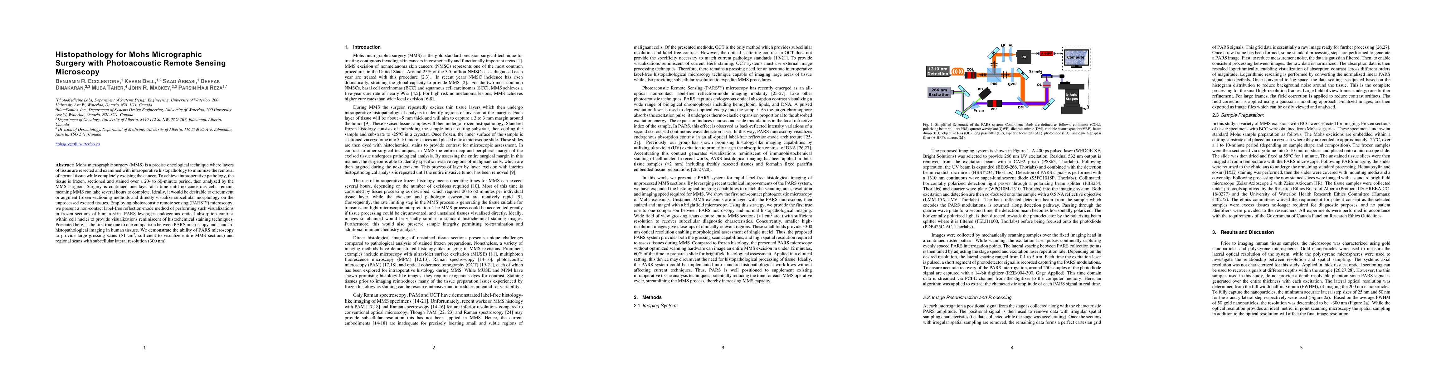
Metrics
AI Quick Summary
Researchers developed a non-contact method using photoacoustic remote sensing microscopy to visualize subcellular morphology in frozen skin tissues without the need for traditional histopathology techniques, promising faster and more accurate Mohs micrographic surgery.
Paper Preview
Abstract
Mohs micrographic surgery (MMS) is a precise oncological technique where layers of tissue are resected and examined with intraoperative histopathology to minimize the removal of normal tissue while completely excising the cancer. To achieve intraoperative pathology, the tissue is frozen, sectioned and stained over a 20- to 60-minute period, then analyzed by the MMS surgeon. Surgery is continued one layer at a time until no cancerous cells remain, meaning MMS can take several hours to complete. Ideally, it would be desirable to circumvent or augment frozen sectioning methods and directly visualize subcellular morphology on the unprocessed excised tissues. Employing photoacoustic remote sensing (PARS) microscopy, we present a non-contact label-free reflection-mode method of performing such visualizations in frozen sections of human skin. PARS leverages endogenous optical absorption contrast within cell nuclei to provide visualizations reminiscent of histochemical staining techniques. Presented here, is the first true one to one comparison between PARS microscopy and standard histopathological imaging in human tissues. We demonstrate the ability of PARS microscopy to provide large grossing scans (>1 cm2, sufficient to visualize entire MMS sections) and regional scans with subcellular lateral resolution (~300 nm).
AI Key Findings
Get AI-generated insights about this paper's methodology, results, significance, and more — seven facets brought into focus.
Impact
Paper Details
PDF Preview
Key Terms
Citation Network
Current paper (gray), citations (green), references (blue)
Display is limited for performance on very large graphs.
Discussion 0