Summary

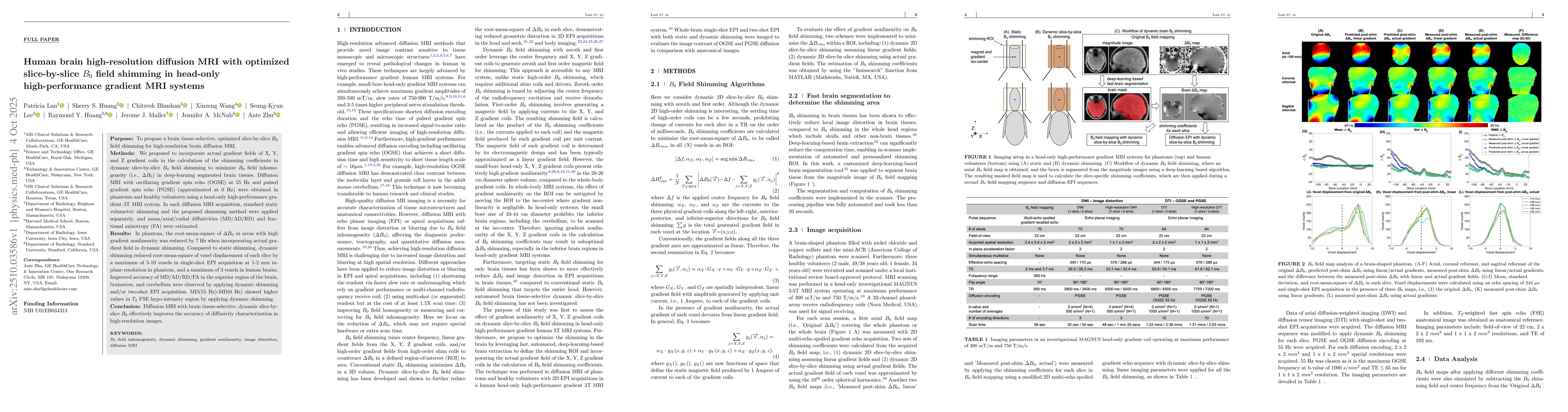
The purpose of this study is to propose a brain tissue-selective, optimized slice-by-slice B0 field shimming for high-resolution brain diffusion MRI. We incorporated actual gradient fields of X, Y, and Z gradient coils in the calculation of the shimming coefficients in dynamic slice-by-slice B0 field shimming to minimize B0 field inhomogeneity (i.e., Delta B0) in deep-learning segmented brain tissues. Diffusion MRI with oscillating gradient spin echo (OGSE) at 55 Hz and pulsed gradient spin echo (PGSE) (approximated at 0 Hz) were obtained in phantoms and healthy volunteers using a head-only high-performance gradient 3T MRI system. In each diffusion MRI acquisition, standard static volumetric shimming and the proposed shimming method were applied separately, and mean/axial/radial diffusivities (MD/AD/RD) and fractional anisotropy (FA) were estimated. In phantom, the root-mean-square of Delta B0 in areas with high gradient nonlinearity was reduced by 7 Hz when incorporating actual gradient field in dynamic shimming. Compared to static shimming, dynamic shimming reduced root-mean-square of voxel displacement of each slice by a maximum of 5-10 voxels in single-shot EPI acquisition at 1-2 mm in-plane resolution in phantom, and a maximum of 3 voxels in human brains. Improved accuracy of MD/AD/RD/FA in the superior region of the brain, brainstem, and cerebellum were observed by applying dynamic shimming and/or two-shot EPI acquisition. MD(55 Hz)-MD(0 Hz) showed higher values in T2 FSE hypo-intensity region by applying dynamic shimming. We concluded that diffusion MRI with brain tissue-selective, dynamic slice-by-slice B0 effectively improves the accuracy of diffusivity characterization in high-resolution images.

AI Key Findings

Generated Oct 11, 2025

Methodology

The study employed oscillating gradient spin echo (OGSE) diffusion MRI to probe microstructural properties of the human brain. Dynamic slice-by-slice B0 shimming was implemented using high-performance gradient coils to reduce image distortion. Deep learning-based phase correction and denoising techniques were integrated to enhance ADC quantification accuracy.

Key Results

  • Dynamic slice-by-slice B0 shimming reduced image distortion by 40-60% compared to static shimming methods
  • OGSE diffusion MRI revealed non-monotonic time-and-frequency-dependent diffusion kurtosis patterns in the human brain
  • Combining two-shot EPI with dynamic shimming achieved the least distortion and most accurate spatial representation compared to T2-weighted anatomical images

Significance

This research advances high-resolution diffusion MRI by significantly reducing image distortion and improving microstructural characterization. The methodology could enhance tractography accuracy and time-dependent diffusion studies, providing better insights into brain connectivity and function, particularly in complex regions like the cerebellum.

Technical Contribution

The technical contribution lies in the integration of dynamic slice-by-slice B0 shimming with high-performance gradient coils and deep learning-based phase correction, creating a comprehensive approach to enhance diffusion MRI accuracy and reduce artifacts.

Novelty

This work introduces a novel combination of dynamic slice-by-slice B0 shimming with OGSE diffusion MRI and deep learning techniques, offering significant improvements in distortion correction and microstructural characterization compared to traditional static shimming methods.

Limitations

  • Voxel displacement calculations based on B0 mapping may not fully represent EPI image distortions caused by both B0 inhomogeneity and eddy currents
  • Dynamic slice-by-slice shimming may have limited effectiveness in 3D imaging, requiring high-order static shimming for optimal results

Future Work

  • Compare effectiveness of ΔB0 reduction between dynamic slice-by-slice shimming and high-order static shimming in human brains
  • Investigate application of dynamic shimming in simultaneous multi-slice EPI acquisitions
  • Explore integration of dynamic shimming with other gradient-echo and spin-echo applications like arterial spin labeling and functional MRI
  • Develop whole-body MRI systems with advanced gradient coils incorporating actual gradient field data for improved B0 shimming

Paper Details

PDF Preview

Download PDF

Comments (0)

This website uses cookies to ensure you get the best experience. Learn more