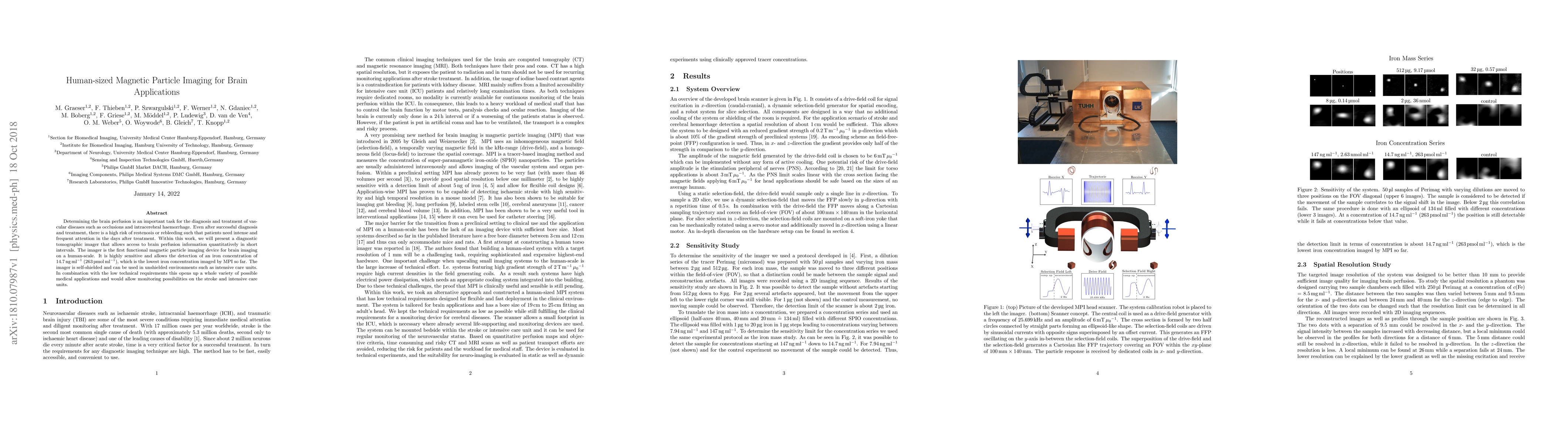
Publication
Metrics
AI Quick Summary
This paper introduces a novel magnetic particle imaging device for human-sized brain imaging, enabling quantitative brain perfusion diagnostics. It is highly sensitive, detecting iron concentrations as low as 14.7 ng/ml, and can operate in unshielded environments, offering potential for monitoring in stroke and intensive care units.
Paper Preview
Abstract
Determining the brain perfusion is an important task for the diagnosis and treatment of vascular diseases such as occlusions and intracerebral haemorrhage. Even after successful diagnosis and treatment, there is a high risk of restenosis or rebleeding such that patients need intense and frequent attention in the days after treatment. Within this work, we will present a diagnostic tomographic imager that allows access to brain perfusion information quantitatively in short intervals. The imager is the first functional magnetic particle imaging device for brain imaging on a human-scale. It is highly sensitive and allows the detection of an iron concentration of 14.7 ng /ml (263 pmol\ml), which is the lowest iron concentration imaged by MPI so far. The imager is self-shielded and can be used in unshielded environments such as intensive care units. In combination with the low technical requirements this opens up a whole variety of possible medical applications and would allow monitoring possibilities on the stroke and intensive care units.
AI Key Findings
Get AI-generated insights about this paper's methodology, results, significance, and more — seven facets brought into focus.
Impact
Paper Details
PDF Preview
Key Terms
Citation Network
Current paper (gray), citations (green), references (blue)
Display is limited for performance on very large graphs.
Discussion 0