01
MethodologyHow they did it
A hybrid 2D and 3D neural network is proposed for liver lesion segmentation
This paper proposes a hybrid cascaded neural network combining 2D and 3D convolutional networks for liver lesion segmentation, achieving a top Dice score of 68.1% on the LiTS challenge. The 2D network segments liver and large tumors, while the 3D network detects small lesions, addressing common segmentation issues.
A hybrid 2D and 3D neural network is proposed for liver lesion segmentation More in Methodology →
Improved performance compared to existing methods — Effective handling of small lesions More in Key Results →
The proposed method has potential applications in medical imaging and tumor detection More in Significance →
Limited by the quality of the LiTS dataset annotations — Potential for over-segmentation or under-segmentation More in Limitations →
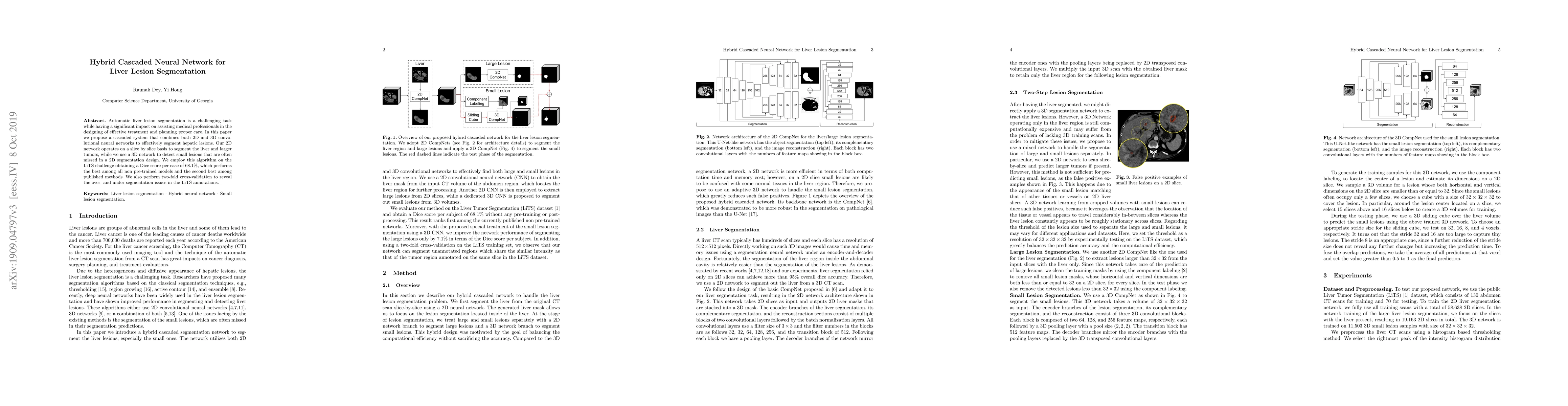
Automatic liver lesion segmentation is a challenging task while having a significant impact on assisting medical professionals in the designing of effective treatment and planning proper care. In this paper we propose a cascaded system that combines both 2D and 3D convolutional neural networks to effectively segment hepatic lesions. Our 2D network operates on a slice by slice basis to segment the liver and larger tumors, while we use a 3D network to detect small lesions that are often missed in a 2D segmentation design. We employ this algorithm on the LiTS challenge obtaining a Dice score per case of 68.1%, which performs the best among all non pre-trained models and the second best among published methods. We also perform two-fold cross-validation to reveal the over- and under-segmentation issues in the LiTS annotations.
Seven facets of this paper, analysed and brought into focus by AI.
The proposed method has potential applications in medical imaging and tumor detection
A hybrid 2D and 3D neural network is proposed for liver lesion segmentation
The proposed method has potential applications in medical imaging and tumor detection
A dedicated 3D segmentation network is designed for small lesions
The proposed method combines 2D and 3D convolutional neural networks for liver lesion segmentation
Current paper (gray), citations (green), references (blue)
Display is limited for performance on very large graphs.
Discussion 0