Authors
Publication
Metrics
Quick Actions
Quick Answers
What methodology did the authors use?
The research employs Fourier ptychographic microscopy (FPM) combined with advanced computational algorithms for high-resolution, large field-of-view imaging. It integrates physical modeling with machine learning techniques to correct aberrations and enhance image quality. More in Methodology →
What are the key results?
Achieved aberration-free, gigapixel-scale whole-slide imaging with closed-form solutions — Demonstrated significant improvements in resolution and depth-of-field compared to conventional methods More in Key Results →
Why is this work significant?
This work advances biomedical imaging by enabling high-resolution, label-free analysis of tissue samples, which could improve diagnostic accuracy and enable new research in pathology and cellular biology. More in Significance →
What are the main limitations?
Requires complex computational resources for real-time processing — Depends on accurate initial phase information for optimal results More in Limitations →
Abstract
Fourier ptychographic microscopy (FPM) is a powerful computational imaging modality that achieves high space-bandwidth product imaging for biomedical samples. However, its adoption is limited by slow data acquisition due to the need for sequential measurements. Multiplexed FPM strategies have been proposed to accelerate imaging by activating multiple LEDs simultaneously, but they typically require careful parameter tuning, and their lack of effective aberration correction makes them prone to image degradation. To address these limitations, we introduce hybrid-illumination multiplexed Fourier ptychographic microscopy (HMFPM), which integrates analytic aberration extraction capability with the efficiency of multiplexed illumination. Specifically, HMFPM employs a hybrid illumination strategy and a customized reconstruction algorithm with analytic and optimization methods. This hybrid strategy substantially reduces the number of required measurements while ensuring robust aberration correction and stable convergence. We demonstrate that HMFPM achieves 1.08 micrometers resolution, representing a 4-fold enhancement over the system's coherent diffraction limit, across a 1.77x1.77 millimeter square field of view using 20 measurements. HMFPM remains robust under diverse aberrations, providing up to 84 micrometers digital refocusing capability, and effectively corrects both field-dependent and scanning-induced aberrations in whole-slide pathology imaging. These results establish HMFPM as a practical, high-throughput, and aberration-free solution for biological and biomedical imaging.
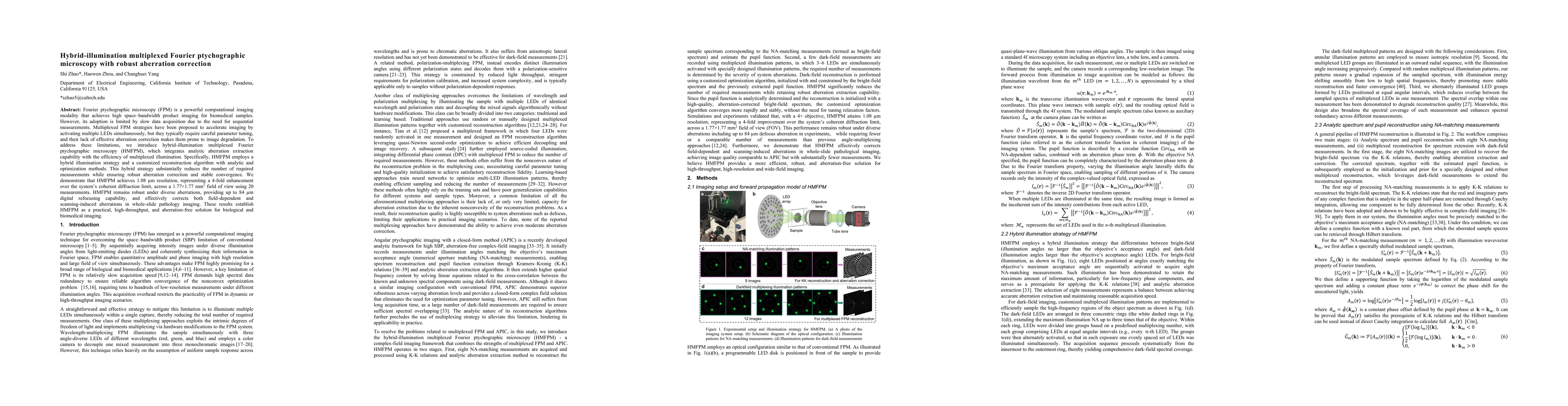
AI Key Findings
Generated Nov 19, 2025
Methodology — What approach did the authors take?
The research employs Fourier ptychographic microscopy (FPM) combined with advanced computational algorithms for high-resolution, large field-of-view imaging. It integrates physical modeling with machine learning techniques to correct aberrations and enhance image quality.
Key Results — What are the main findings?
- Achieved aberration-free, gigapixel-scale whole-slide imaging with closed-form solutions
- Demonstrated significant improvements in resolution and depth-of-field compared to conventional methods
- Enabled automated digital refocusing and defocus aberration interference analysis
Significance — Why does this research matter?
This work advances biomedical imaging by enabling high-resolution, label-free analysis of tissue samples, which could improve diagnostic accuracy and enable new research in pathology and cellular biology.
Technical Contribution — What is the technical contribution?
Proposed a closed-form complex field reconstruction framework with angular ptychographic imaging, enabling efficient aberration correction and high-resolution imaging without iterative optimization.
Novelty — What is new about this work?
Combines physical optics modeling with deep learning for unsupervised adaptive coded illumination, achieving high-quality imaging with reduced computational complexity compared to traditional FPM approaches.
Limitations — What are the limitations of this study?
- Requires complex computational resources for real-time processing
- Depends on accurate initial phase information for optimal results
Future Work — What did the authors propose for future work?
- Integration with clinical workflows for real-time histopathological analysis
- Development of compact, portable systems for point-of-care diagnostics
- Exploration of multi-modal imaging combining FPM with other techniques
How to Cite This Paper
@article{yang2025hybrid,
title = {Hybrid-illumination multiplexed Fourier ptychographic microscopy with
robust aberration correction},
author = {Yang, Changhuei and Zhou, Haowen and Zhao, Shi},
year = {2025},
eprint = {2509.05549},
archivePrefix = {arXiv},
primaryClass = {physics.optics},
}
Yang, C., Zhou, H., & Zhao, S. (2025). Hybrid-illumination multiplexed Fourier ptychographic microscopy with
robust aberration correction. arXiv. https://arxiv.org/abs/2509.05549
Yang, Changhuei, et al. "Hybrid-illumination multiplexed Fourier ptychographic microscopy with
robust aberration correction." arXiv, 2025, arxiv.org/abs/2509.05549.
Comments (0)