HydraViT: Adaptive Multi-Branch Transformer for Multi-Label Disease Classification from Chest X-ray Images
Publication
Metrics
AI Quick Summary
HydraViT combines a transformer backbone with a multi-branch output module to enhance multi-label disease classification from chest X-ray images, outperforming traditional CNNs and attention-guided methods by leveraging long-range context and adaptive focus on critical regions.
Paper Preview
Abstract
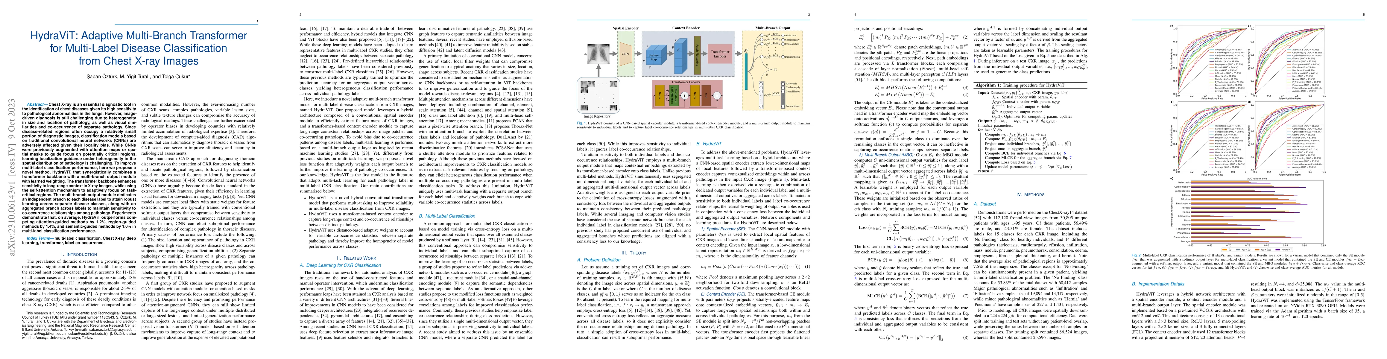
Chest X-ray is an essential diagnostic tool in the identification of chest diseases given its high sensitivity to pathological abnormalities in the lungs. However, image-driven diagnosis is still challenging due to heterogeneity in size and location of pathology, as well as visual similarities and co-occurrence of separate pathology. Since disease-related regions often occupy a relatively small portion of diagnostic images, classification models based on traditional convolutional neural networks (CNNs) are adversely affected given their locality bias. While CNNs were previously augmented with attention maps or spatial masks to guide focus on potentially critical regions, learning localization guidance under heterogeneity in the spatial distribution of pathology is challenging. To improve multi-label classification performance, here we propose a novel method, HydraViT, that synergistically combines a transformer backbone with a multi-branch output module with learned weighting. The transformer backbone enhances sensitivity to long-range context in X-ray images, while using the self-attention mechanism to adaptively focus on task-critical regions. The multi-branch output module dedicates an independent branch to each disease label to attain robust learning across separate disease classes, along with an aggregated branch across labels to maintain sensitivity to co-occurrence relationships among pathology. Experiments demonstrate that, on average, HydraViT outperforms competing attention-guided methods by 1.2%, region-guided methods by 1.4%, and semantic-guided methods by 1.0% in multi-label classification performance.
AI Key Findings
Get AI-generated insights about this paper's methodology, results, significance, and more — seven facets brought into focus.
Impact
Paper Details
Authors
PDF Preview
Key Terms
Citation Network
Current paper (gray), citations (green), references (blue)
Display is limited for performance on very large graphs.
Discussion 0