Publication
Metrics
AI Quick Summary
This paper uses cryogenic transmission electron microscopy to directly image the localization of excitons within a moiré unit-cell in WS2 and WSe2 monolayers, revealing that the exciton wavefunction is confined to a ~2 nm radius around the highest-energy stacking site, forming a triangular lattice. The findings suggest that atomic reconstructions create strong moiré potentials, paving the way for engineering strain to develop new excitonic lattices.
Paper Preview
Abstract
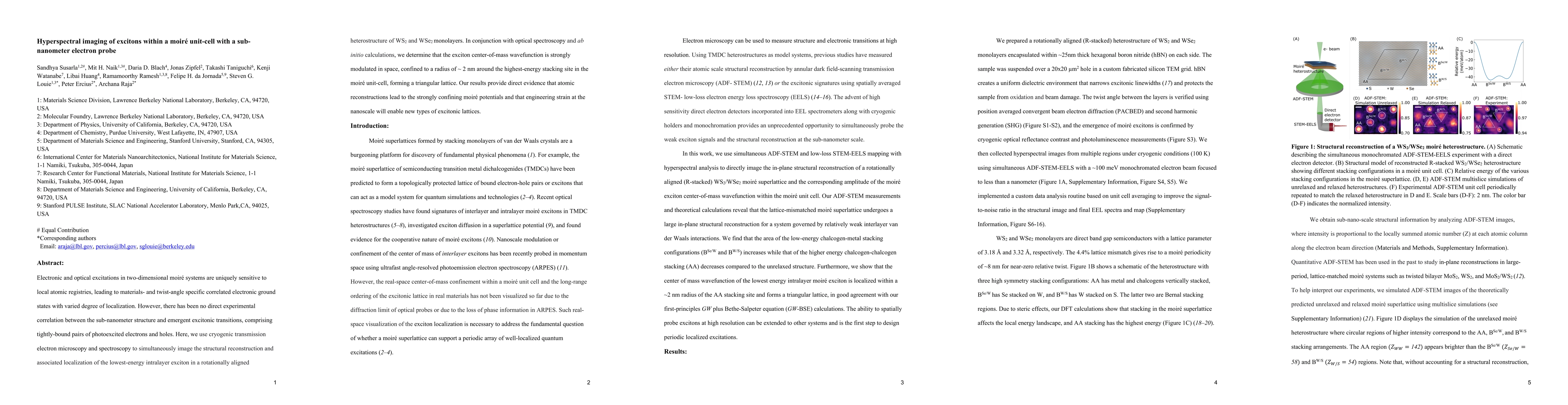
Electronic and optical excitations in two-dimensional moir\'e systems are uniquely sensitive to local atomic registries, leading to materials- and twist-angle specific correlated electronic ground states with varied degree of localization. However, there has been no direct experimental correlation between the sub-nanometer structure and emergent excitonic transitions, comprising tightly-bound pairs of photoexcited electrons and holes. Here, we use cryogenic transmission electron microscopy and spectroscopy to simultaneously image the structural reconstruction and associated localization of the lowest-energy intralayer exciton in a rotationally aligned heterostructure of WS2 and WSe2 monolayers. In conjunction with optical spectroscopy and ab initio calculations, we determine that the exciton center-of-mass wavefunction is strongly modulated in space, confined to a radius of ~ 2 nm around the highest-energy stacking site in the moir\'e unit-cell, forming a triangular lattice. Our results provide direct evidence that atomic reconstructions lead to the strongly confining moir\'e potentials and that engineering strain at the nanoscale will enable new types of excitonic lattices.
AI Key Findings
Get AI-generated insights about this paper's methodology, results, significance, and more — seven facets brought into focus.
Impact
Paper Details
Authors
PDF Preview
Key Terms
Citation Network
Current paper (gray), citations (green), references (blue)
Display is limited for performance on very large graphs.
Discussion 0