Publication
Metrics
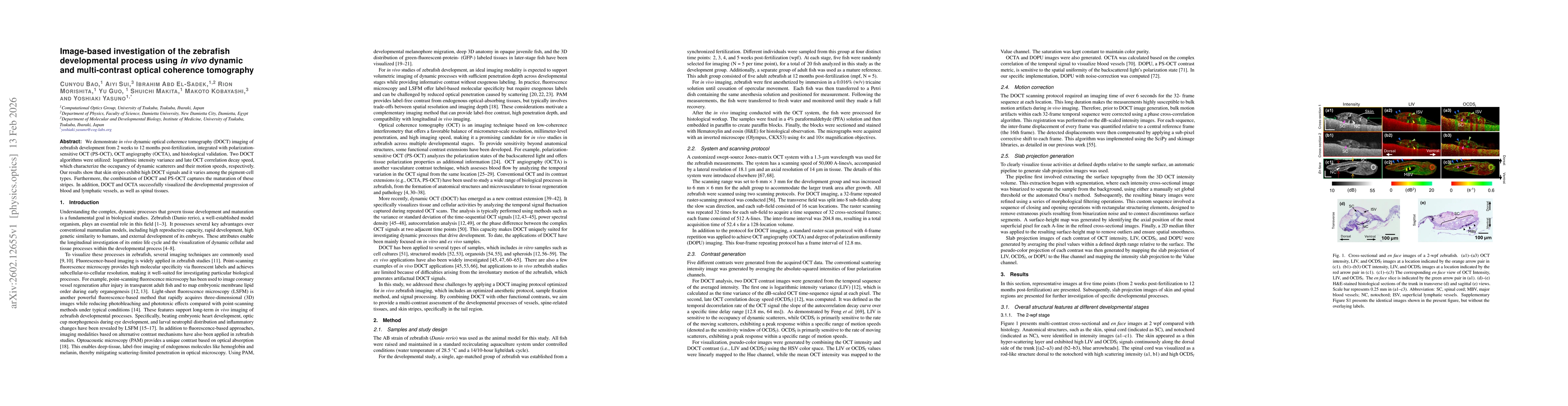
Paper Preview
Abstract
We demonstrate in vivo dynamic optical coherence tomography (DOCT) imaging of zebrafish development from 2 weeks to 12 months post-fertilization, integrated with polarization-sensitive OCT (PS-OCT), OCT angiography (OCTA), and histological validation. Two DOCT algorithms were utilized: logarithmic intensity variance and late OCT correlation decay speed, which characterize the occupancy of dynamic scatterers and their motion speeds, respectively. Our results show that skin stripes exhibit high DOCT signals and it varies among the pigment-cell types. Furthermore, the combination of DOCT and PS-OCT captures the maturation of these stripes. In addition, DOCT and OCTA successfully visualized the developmental progression of blood and lymphatic vessels, as well as spinal tissues.
AI Key Findings
Get AI-generated insights about this paper's methodology, results, significance, and more — seven facets brought into focus.
Discussion 0