Image enhancement in acoustic-resolution photoacoustic microscopy enabled by a novel directional algorithm
Publication
Metrics
AI Quick Summary
A new algorithm enhances image resolution and signal-to-noise ratio in acoustic-resolution photoacoustic microscopy, allowing for high-quality imaging of microvasculature with a depth of focus of 1.8 mm.
Paper Preview
Abstract
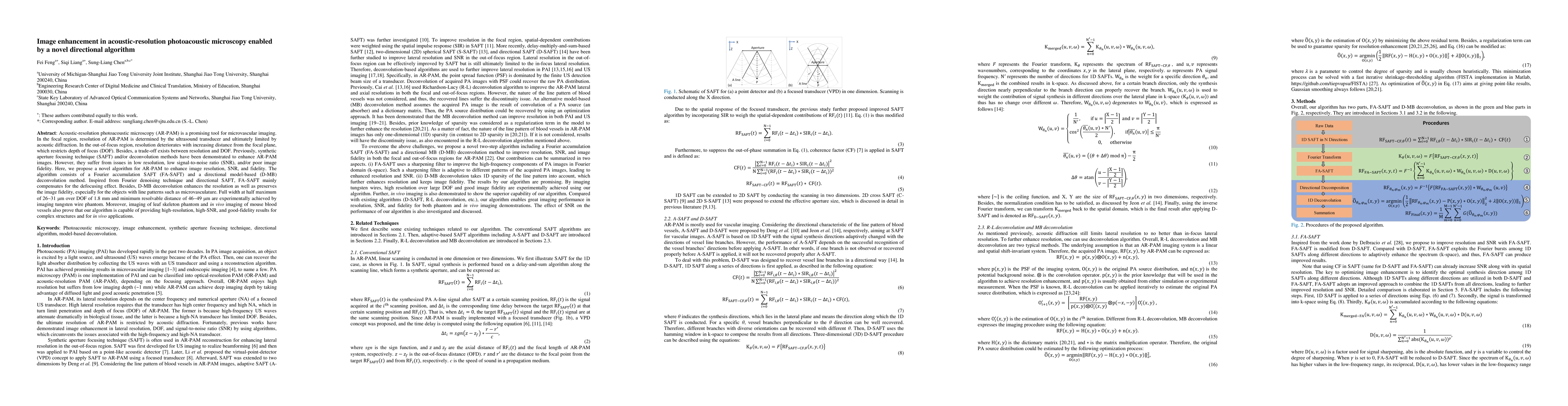
Acoustic-resolution photoacoustic microscopy (AR-PAM) is a promising tool for microvascular imaging. In the focal region, resolution of AR-PAM is determined by the ultrasound transducer and ultimately limited by acoustic diffraction. In the out-of-focus region, resolution deteriorates with increasing distance from the focal plane, which restricts depth of focus (DOF). Besides, a trade-off exists between resolution and DOF. Previously, synthetic aperture focusing technique (SAFT) and/or deconvolution methods have been demonstrated to enhance AR-PAM images. However, they suffer from issues in low resolution, low signal-to-noise ratio (SNR), and/or poor image fidelity. Here, we propose a novel algorithm for AR-PAM to enhance image resolution, SNR, and fidelity. The algorithm consists of a Fourier accumulation SAFT (FA-SAFT) and a directional model-based (D-MB) deconvolution method. Inspired from Fourier denoising technique and directional SAFT, FA-SAFT mainly compensates for the defocusing effect. Besides, D-MB deconvolution enhances the resolution as well as preserves the image fidelity, especially for the objects with line patterns such as microvasculature. Full width at half maximum of 26-31 um over DOF of 1.8 mm and minimum resolvable distance of 46-49 um are experimentally achieved by imaging tungsten wire phantom. Moreover, imaging of leaf skeleton phantom and in vivo imaging of mouse blood vessels also prove that our algorithm is capable of providing high-resolution, high-SNR, and good-fidelity results for complex structures and for in vivo applications.
AI Key Findings
Get AI-generated insights about this paper's methodology, results, significance, and more — seven facets brought into focus.
Impact
Paper Details
Authors
PDF Preview
Key Terms
Citation Network
Current paper (gray), citations (green), references (blue)
Display is limited for performance on very large graphs.
Discussion 0