Image Segmentation of Zona-Ablated Human Blastocysts
Publication
Metrics
AI Quick Summary
A deep learning-based method improves the accuracy of measuring blastocyst expansion in human embryos, with potential to increase IVF success rates by detecting genetic abnormalities.
Paper Preview
Abstract
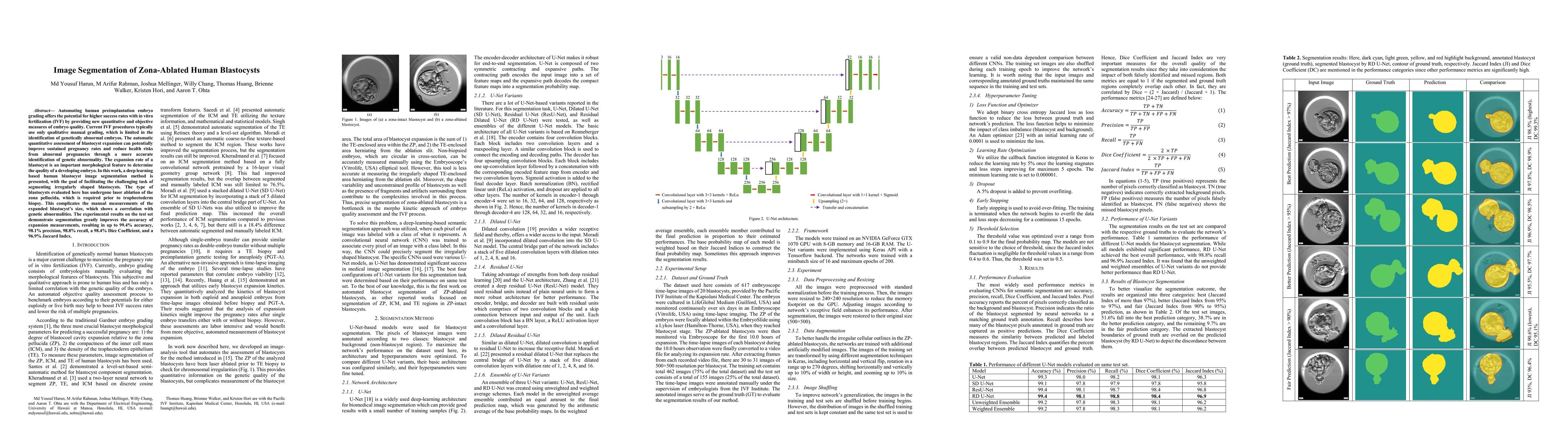
Automating human preimplantation embryo grading offers the potential for higher success rates with in vitro fertilization (IVF) by providing new quantitative and objective measures of embryo quality. Current IVF procedures typically use only qualitative manual grading, which is limited in the identification of genetically abnormal embryos. The automatic quantitative assessment of blastocyst expansion can potentially improve sustained pregnancy rates and reduce health risks from abnormal pregnancies through a more accurate identification of genetic abnormality. The expansion rate of a blastocyst is an important morphological feature to determine the quality of a developing embryo. In this work, a deep learning based human blastocyst image segmentation method is presented, with the goal of facilitating the challenging task of segmenting irregularly shaped blastocysts. The type of blastocysts evaluated here has undergone laser ablation of the zona pellucida, which is required prior to trophectoderm biopsy. This complicates the manual measurements of the expanded blastocyst's size, which shows a correlation with genetic abnormalities. The experimental results on the test set demonstrate segmentation greatly improves the accuracy of expansion measurements, resulting in up to 99.4% accuracy, 98.1% precision, 98.8% recall, a 98.4% Dice Coefficient, and a 96.9% Jaccard Index.
AI Key Findings
Get AI-generated insights about this paper's methodology, results, significance, and more — seven facets brought into focus.
Impact
Paper Details
Authors
PDF Preview
Key Terms
Citation Network
Current paper (gray), citations (green), references (blue)
Display is limited for performance on very large graphs.
Discussion 0