Publication
Metrics
AI Quick Summary
This study employs low-energy electron microscopy in mirror mode to achieve submicron resolution imaging of ferroelectric domains in bismuth ferrite, demonstrating its potential for rapid, full-field domain visualization compared to piezoresponse force microscopy.
Paper Preview
Abstract
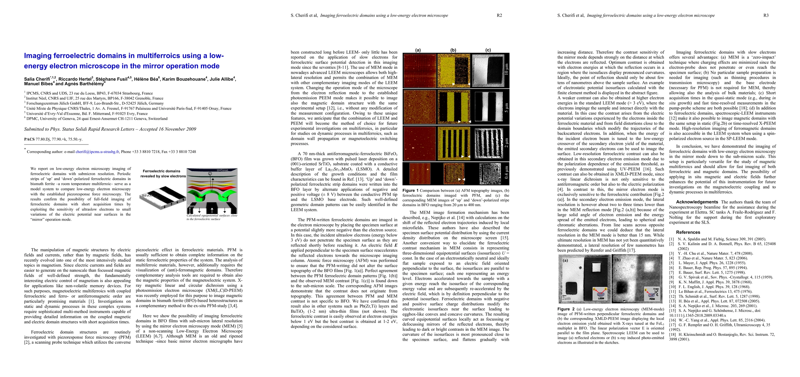
We report on low-energy electron microscopy imaging of ferroelectric domains with submicron resolution. Periodic strips of 'up' and 'down'-polarized ferroelectric domains in bismuth ferrite -a room temperature multiferroic- serve as a model system to compare low-energy electron microscopy with the established piezoresponse force microscopy. The results confirm the possibility of full-field imaging of ferroelectric domains with short acquisition times by exploiting the sensitivity of ultraslow electrons to small variations of the electric potential near surfaces in the "mirror" operation mode.
AI Key Findings
Get AI-generated insights about this paper's methodology, results, significance, and more — seven facets brought into focus.
Impact
Paper Details
PDF Preview
Key Terms
Citation Network
Current paper (gray), citations (green), references (blue)
Display is limited for performance on very large graphs.
Discussion 0