AI Quick Summary
This study employs charge gradient microscopy enhanced by principal component analysis to image ferroelectric domains in lithium niobate, revealing domain structures and polarization dynamics. The method captures dynamic polarization changes and surface charge distributions, providing quantitative insights into ferroelectric domain behavior.
Paper Preview
Abstract
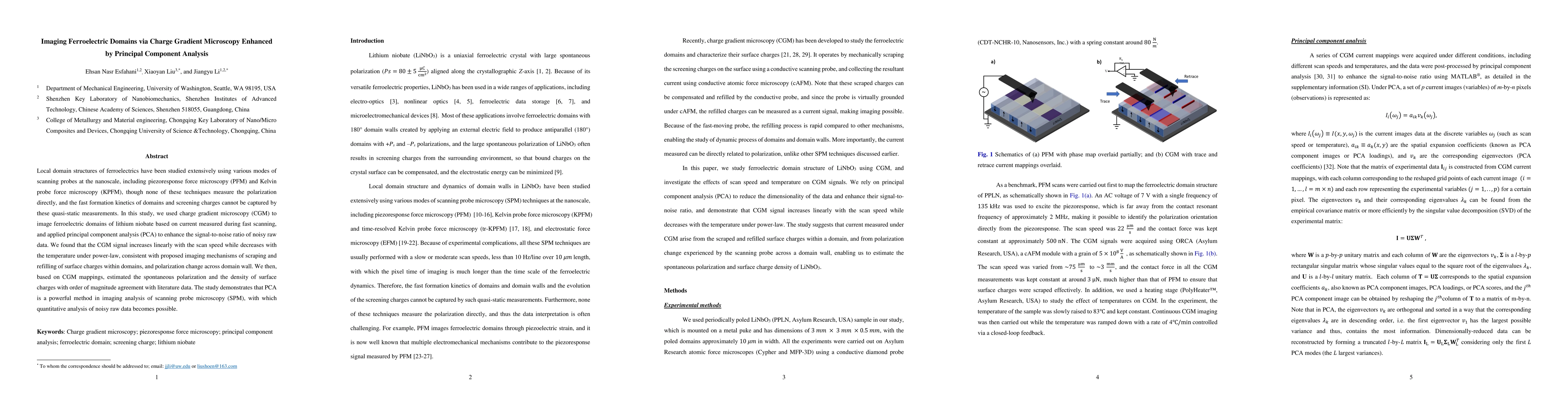
Local domain structures of ferroelectrics have been studied extensively using various modes of scanning probes at the nanoscale, including piezoresponse force microscopy (PFM) and Kelvin probe force microscopy (KPFM), though none of these techniques measure the polarization directly, and the fast formation kinetics of domains and screening charges cannot be captured by these quasi-static measurements. In this study, we used charge gradient microscopy (CGM) to image ferroelectric domains of lithium niobate based on current measured during fast scanning, and applied principal component analysis (PCA) to enhance the signal-to-noise ratio of noisy raw data. We found that the CGM signal increases linearly with the scan speed while decreases with the temperature under power-law, consistent with proposed imaging mechanisms of scraping and refilling of surface charges within domains, and polarization change across domain wall. We then, based on CGM mappings, estimated the spontaneous polarization and the density of surface charges with order of magnitude agreement with literature data. The study demonstrates that PCA is a powerful method in imaging analysis of scanning probe microscopy (SPM), with which quantitative analysis of noisy raw data becomes possible.
AI Key Findings
Get AI-generated insights about this paper's methodology, results, and significance.
Paper Details
How to Cite This Paper
@article{anon2017imaging,
  title         = {Imaging Ferroelectric Domains via Charge Gradient Microscopy Enhanced by
  Principal Component Analysis},
  year          = {2017},
  eprint        = {1706.02345},
  archivePrefix = {arXiv},
  primaryClass  = {cond-mat.mtrl-sci},
  doi           = {10.1016/j.jmat.2017.07.001},
}(2017). Imaging Ferroelectric Domains via Charge Gradient Microscopy Enhanced by
Principal Component Analysis. arXiv. https://doi.org/10.1016/j.jmat.2017.07.001"Imaging Ferroelectric Domains via Charge Gradient Microscopy Enhanced by
Principal Component Analysis." arXiv, 2017, doi.org/10.1016/j.jmat.2017.07.001.PDF Preview
Key Terms
Citation Network
Current paper (gray), citations (green), references (blue)
Display is limited for performance on very large graphs.
Similar Papers
Found 4 papersHigh-contrast imaging of 180{\deg} ferroelectric domains by optical microscopy using ferroelectric liquid crystals
Xavier Moya, Tim D. Wilkinson, Jan P. F. Lagerwall et al.
Imaging Ferroelectrics: Charge Gradient Microscopy (CGM) versus Potential Gradient Microscopy (PGM)
Hamza Waseem, Jesi R. Maguire, Amit Kumar et al.
Principal Component Analysis in Application to Brillouin Microscopy Data
Hadi Mahmodi, Christopher G. Poulton, Mathew N. Lesley et al.
Fast and Provable Tensor Robust Principal Component Analysis via Scaled Gradient Descent
Tian Tong, Harry Dong, Cong Ma et al.
| Title | Authors | Year | Actions |
|---|
Comments (0)