Summary
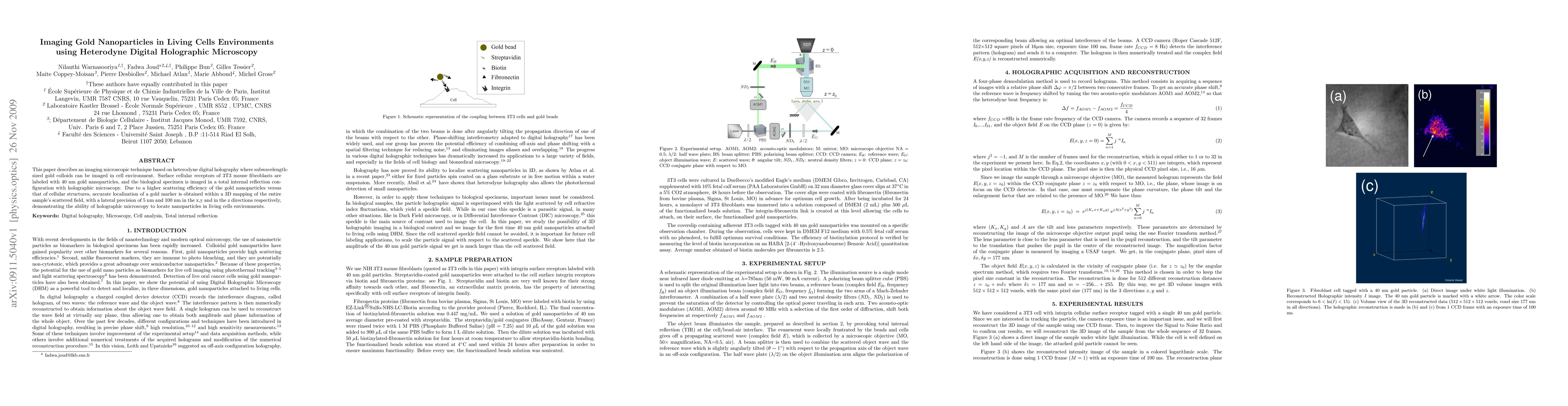
This paper describes an imaging microscopic technique based on heterodyne digital holography where subwavelength-sized gold colloids can be imaged in cell environment. Surface cellular receptors of 3T3 mouse fibroblasts are labeled with 40 nm gold nanoparticles, and the biological specimen is imaged in a total internal reflection configuration with holographic microscopy. Due to a higher scattering efficiency of the gold nanoparticles versus that of cellular structures, accurate localization of a gold marker is obtained within a 3D mapping of the entire sample's scattered field, with a lateral precision of 5 nm and 100 nm in the x,y and in the z directions respectively, demonstrating the ability of holographic microscopy to locate nanoparticles in living cells environments.
AI Key Findings
Get AI-generated insights about this paper's methodology, results, and significance.
Paper Details
PDF Preview
Key Terms
Citation Network
Current paper (gray), citations (green), references (blue)
Display is limited for performance on very large graphs.
Similar Papers
Found 4 papersCorrelative light electron microscopy using small gold nanoparticles as single probes
Wolfgang Langbein, Lukas Payne, Paola Borri et al.
| Title | Authors | Year | Actions |
|---|
Comments (0)