Imaging mesoscopic nuclear spin noise with a diamond magnetometer

Source: ArXiv

AI Quick Summary

Researchers develop a diamond magnetometer to detect nanoscale nuclear spin noise for high-resolution imaging and spectroscopy of biological samples. The sensor can probe fluctuations in classical dipolar fields, allowing for detailed sample spectra and image contrast.

Share: Share on X LinkedIn

Summary

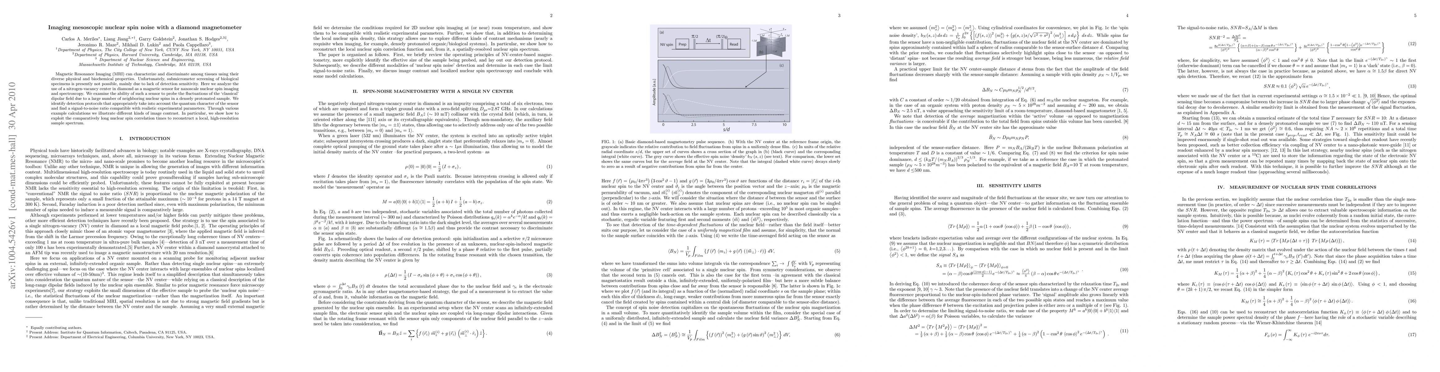
Magnetic Resonance Imaging (MRI) can characterize and discriminate among tissues using their diverse physical and biochemical properties. Unfortunately, submicrometer screening of biological specimens is presently not possible, mainly due to lack of detection sensitivity. Here we analyze the use of a nitrogen-vacancy center in diamond as a magnetic sensor for nanoscale nuclear spin imaging and spectroscopy. We examine the ability of such a sensor to probe the fluctuations of the "classical" dipolar field due to a large number of neighboring nuclear spins in a densely protonated sample. We identify detection protocols that appropriately take into account the quantum character of the sensor and find a signal-to-noise ratio compatible with realistic experimental parameters. Through various example calculations we illustrate different kinds of image contrast. In particular, we show how to exploit the comparatively long nuclear spin correlation times to reconstruct a local, high-resolution sample spectrum.

AI Key Findings

Generated Sep 07, 2025

Methodology

The research uses a nitrogen-vacancy (NV) center in diamond as a magnetic sensor for nanoscale nuclear spin imaging and spectroscopy, examining its ability to probe fluctuations of nuclear spin fields in densely protonated samples.

Key Results

  • Identified detection protocols that consider the quantum nature of the NV center sensor and achieve a signal-to-noise ratio compatible with realistic experimental parameters.
  • Demonstrated methods to create various kinds of image contrast, including reconstructing a local, high-resolution sample spectrum using comparatively long nuclear spin correlation times.
  • Established a criterion for spin noise dominance, d ≤ (kBT/(mNBA))^2/3 ρ^-1/3_N, for practical sensor-sample distances.

Significance

This work presents a novel approach for nanoscale magnetic resonance imaging (MRI), potentially overcoming current limitations of submicrometer screening due to lack of detection sensitivity, and opening new avenues for studying biological specimens at unprecedented resolution.

Technical Contribution

The paper presents analytical and numerical calculations demonstrating the use of NV centers in diamond for detecting and imaging nuclear spin noise with nanoscale resolution, providing protocols and methods for signal extraction and image reconstruction.

Novelty

This research introduces a new method for nanoscale magnetic resonance imaging using NV centers in diamond, which circumvents limitations of conventional MRI by enabling detection of magnetic field fluctuations from individual nuclear spins, thus paving the way for high-resolution imaging of biological specimens.

Limitations

  • The current sensitivity limit could be improved significantly with the availability of single-shot readout techniques.
  • Practical implementation may face challenges in achieving and maintaining the required coherence times for high-resolution imaging.

Future Work

  • Explore and develop single-shot readout strategies, such as enhanced collection efficiency via coupling to nano-photonic waveguides or readout enhanced by nuclear spin memory.
  • Investigate the application of this technique to diverse biological systems to demonstrate its versatility and potential for real-world impact.

Paper Details

PDF Preview

Download PDF

Key Terms

sensor (0.276)
nuclear (0.243)
imaging (0.184)
nitrogen (0.158)
detection (0.155)
dipolar (0.154)
sample (0.153)
spin (0.147)
Title Authors Year Actions

Comments (0)

This website uses cookies to ensure you get the best experience. Learn more