Imaging of lateral spin valves with soft X-ray microscopy
Publication
Metrics
AI Quick Summary
This finding suggests that existing methods may not be effective in directly imaging spin accumulation in materials like Cu during illumination with circular polarized x-rays.
Paper Preview
Abstract
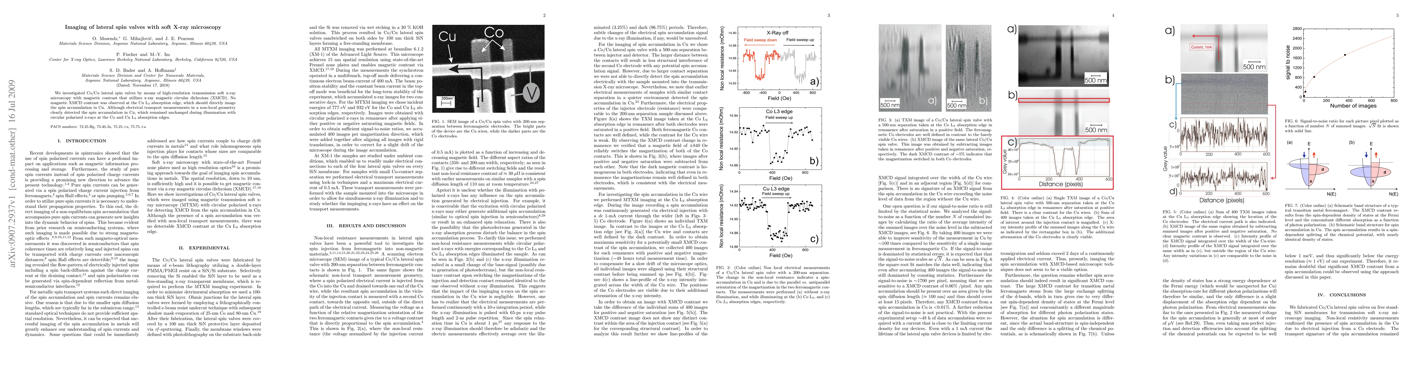
We investigated Co/Cu lateral spin valves by means of high-resolution transmission soft x-ray microscopy with magnetic contrast that utilizes x-ray magnetic circular dichroism (XMCD). No magnetic XMCD contrast was observed at the Cu L$_3$ absorption edge, which should directly image the spin accumulation in Cu. Although electrical transport measurements in a non-local geometry clearly detected the spin accumulation in Cu, which remained unchanged during illumination with circular polarized x-rays at the Co and Cu L$_3$ absorption edges.
AI Key Findings
Get AI-generated insights about this paper's methodology, results, significance, and more — seven facets brought into focus.
Impact
Paper Details
PDF Preview
Key Terms
Citation Network
Current paper (gray), citations (green), references (blue)
Display is limited for performance on very large graphs.
Discussion 0