Summary
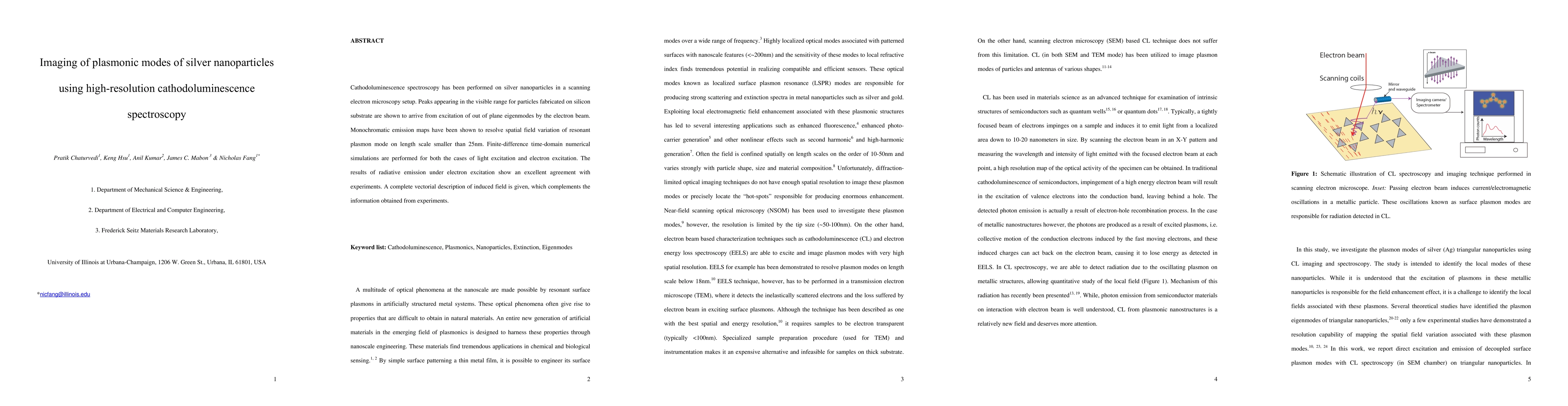
Cathodoluminescence spectroscopy has been performed on silver nanoparticles in a scanning electron microscopy setup. Peaks appearing in the visible range for particles fabricated on silicon substrate are shown to arrive from excitation of out of plane eigenmodes by the electron beam. Monochromatic emission maps have been shown to resolve spatial field variation of resonant plasmon mode on length scale smaller than 25nm. Finite-difference time-domain numerical simulations are performed for both the cases of light excitation and electron excitation. The results of radiative emission under electron excitation show an excellent agreement with experiments. A complete vectorial description of induced field is given, which complements the information obtained from experiments.
AI Key Findings
Get AI-generated insights about this paper's methodology, results, and significance.
Paper Details
PDF Preview
Key Terms
Citation Network
Current paper (gray), citations (green), references (blue)
Display is limited for performance on very large graphs.
Similar Papers
Found 4 papersExcitation and Imaging of Resonant Optical Modes of Au Triangular Nano-Antennas Using Cathodoluminescence Spectroscopy
Anil Kumar, Edmond Chow, Nicholas X. Fang et al.
Cathodoluminescence excitation spectroscopy: nanoscale imaging of excitation pathways
Takashi Taniguchi, Kenji Watanabe, Steffi Y. Woo et al.
| Title | Authors | Year | Actions |
|---|
Comments (0)