Summary
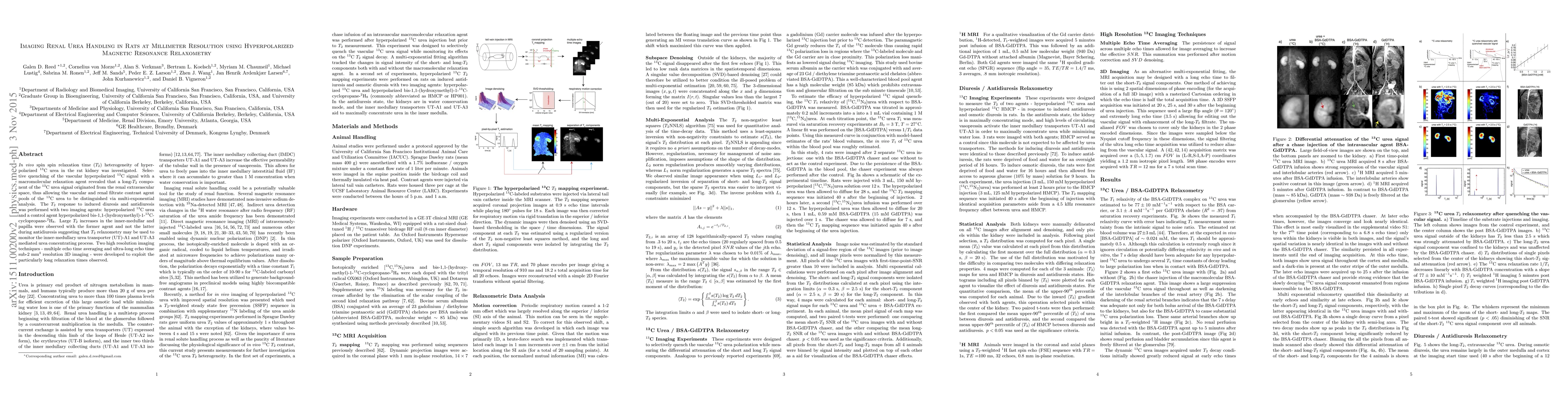
\textit{In vivo} spin spin relaxation time ($T_2$) heterogeneity of hyperpolarized \textsuperscript{13}C urea in the rat kidney was investigated. Selective quenching of the vascular hyperpolarized \textsuperscript{13}C signal with a macromolecular relaxation agent revealed that a long-$T_2$ component of the \textsuperscript{13}C urea signal originated from the renal extravascular space, thus allowing the vascular and renal filtrate contrast agent pools of the \textsuperscript{13}C urea to be distinguished via multi-exponential analysis. The $T_2$ response to induced diuresis and antidiuresis was performed with two imaging agents: hyperpolarized \textsuperscript{13}C urea and a control agent hyperpolarized bis-1,1-(hydroxymethyl)-1-\textsuperscript{13}C-cyclopropane-$^2\textrm{H}_8$. Large $T_2$ increases in the inner-medullar and papilla were observed with the former agent and not the latter during antidiuresis suggesting that $T_2$ relaxometry may be used to monitor the inner-medullary urea transporter (UT)-A1 and UT-A3 mediated urea concentrating process. Two high resolution imaging techniques - multiple echo time averaging and ultra-long echo time sub-2 mm$^3$ resolution 3D imaging - were developed to exploit the particularly long relaxation times observed.
AI Key Findings
Get AI-generated insights about this paper's methodology, results, and significance.
Paper Details
PDF Preview
Key Terms
Citation Network
Current paper (gray), citations (green), references (blue)
Display is limited for performance on very large graphs.
Similar Papers
Found 4 papersRelaxometry Guided Quantitative Cardiac Magnetic Resonance Image Reconstruction
Yi Zhang, Yidong Zhao, Qian Tao
| Title | Authors | Year | Actions |
|---|
Comments (0)