Publication
Metrics
AI Quick Summary
This paper introduces a new imaging technique combining high-resolution optically-sectioned images with low-resolution whole-heart recordings to capture the full spatio-temporal dynamics of the beating embryonic heart. The method uses phase stamping to reconstruct the 3D beating heart, demonstrated effectively in zebrafish embryos.
Paper Preview
Abstract
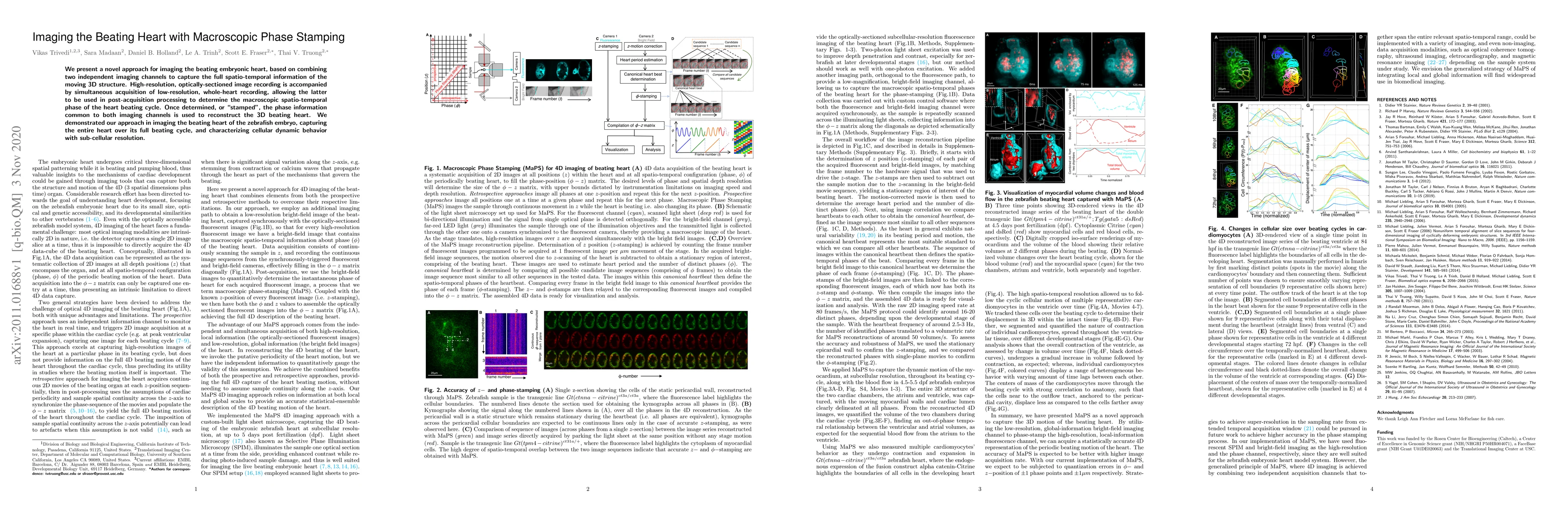
We present a novel approach for imaging the beating embryonic heart, based on combining two independent imaging channels to capture the full spatio-temporal information of the moving 3D structure. High-resolution, optically-sectioned image recording is accompanied by simultaneous acquisition of low-resolution, whole-heart recording, allowing the latter to be used in post-acquisition processing to determine the macroscopic spatio-temporal phase of the heart beating cycle. Once determined, or 'stamped', the phase information common to both imaging channels is used to reconstruct the 3D beating heart. We demonstrated our approach in imaging the beating heart of the zebrafish embryo, capturing the entire heart over its full beating cycle, and characterizing cellular dynamic behavior with sub-cellular resolution.
AI Key Findings
Get AI-generated insights about this paper's methodology, results, significance, and more — seven facets brought into focus.
Impact
Paper Details
Authors
PDF Preview
Key Terms
Citation Network
Current paper (gray), citations (green), references (blue)
Display is limited for performance on very large graphs.
Discussion 0