Quick Answers

What methodology did the authors use?

The study employed a transfer learning approach using pre-trained models (HistoPLUS and EpiSeg) to segment and detect cellular structures in histopathological images from IBD datasets. The models were validated on external cohorts (FINBB and IBDColEpis) to assess their performance across different staining protocols and tissue samples. More in Methodology →

What are the key results?

HistoPLUS achieved high panoptic quality (0.605 on HistoVAL, 0.586 on SPARCIBD) with strong detection (0.753) and segmentation (0.801) metrics on oncology validation data. — EpiSeg demonstrated high precision-recall performance on the IBD-ColEpitest set, with average precision metrics indicating reliable epithelium detection. More in Key Results →

Why is this work significant?

This research advances automated histopathological analysis for IBD, enabling more efficient and objective assessment of tissue samples. The results suggest potential for deploying AI tools in clinical settings to support diagnosis and monitor treatment response. More in Significance →

What are the main limitations?

Performance metrics may vary with different staining protocols and tissue types not included in the study. — The study primarily focused on IBD datasets, limiting generalizability to other diseases. More in Limitations →

Paper Preview

Abstract

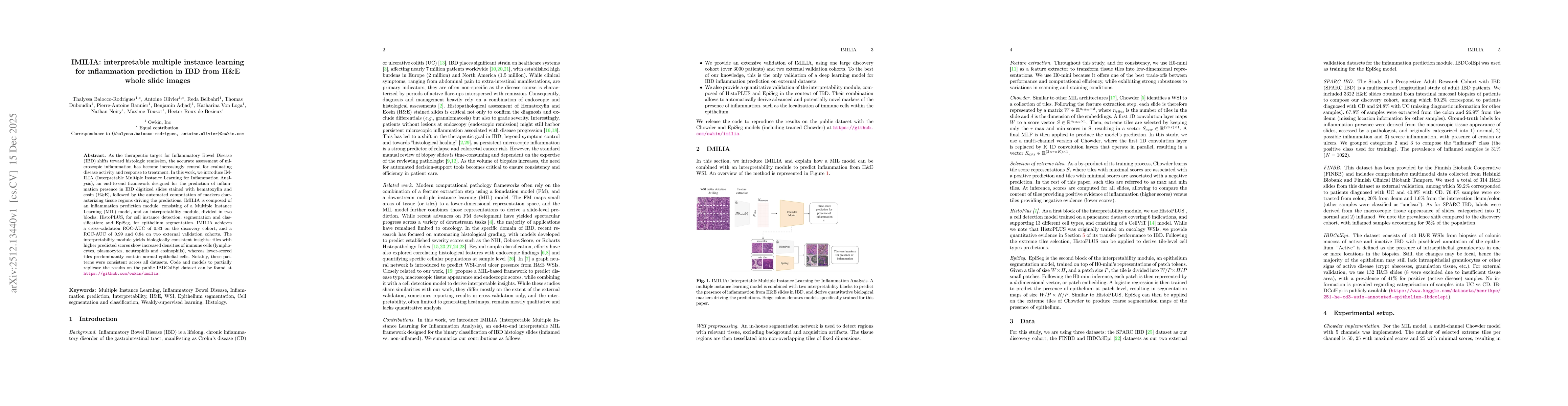
As the therapeutic target for Inflammatory Bowel Disease (IBD) shifts toward histologic remission, the accurate assessment of microscopic inflammation has become increasingly central for evaluating disease activity and response to treatment. In this work, we introduce IMILIA (Interpretable Multiple Instance Learning for Inflammation Analysis), an end-to-end framework designed for the prediction of inflammation presence in IBD digitized slides stained with hematoxylin and eosin (H&E), followed by the automated computation of markers characterizing tissue regions driving the predictions. IMILIA is composed of an inflammation prediction module, consisting of a Multiple Instance Learning (MIL) model, and an interpretability module, divided in two blocks: HistoPLUS, for cell instance detection, segmentation and classification; and EpiSeg, for epithelium segmentation. IMILIA achieves a cross-validation ROC-AUC of 0.83 on the discovery cohort, and a ROC-AUC of 0.99 and 0.84 on two external validation cohorts. The interpretability module yields biologically consistent insights: tiles with higher predicted scores show increased densities of immune cells (lymphocytes, plasmocytes, neutrophils and eosinophils), whereas lower-scored tiles predominantly contain normal epithelial cells. Notably, these patterns were consistent across all datasets. Code and models to partially replicate the results on the public IBDColEpi dataset can be found at https://github.com/owkin/imilia.

AI Key Findings

Generated Dec 16, 2025

Methodology — What approach did the authors take?

The study employed a transfer learning approach using pre-trained models (HistoPLUS and EpiSeg) to segment and detect cellular structures in histopathological images from IBD datasets. The models were validated on external cohorts (FINBB and IBDColEpis) to assess their performance across different staining protocols and tissue samples.

Key Results — What are the main findings?

  • HistoPLUS achieved high panoptic quality (0.605 on HistoVAL, 0.586 on SPARCIBD) with strong detection (0.753) and segmentation (0.801) metrics on oncology validation data.
  • EpiSeg demonstrated high precision-recall performance on the IBD-ColEpitest set, with average precision metrics indicating reliable epithelium detection.
  • The models showed consistent performance across external validation cohorts, with minimal staining distribution shifts observed between discovery and validation datasets.

Significance — Why does this research matter?

This research advances automated histopathological analysis for IBD, enabling more efficient and objective assessment of tissue samples. The results suggest potential for deploying AI tools in clinical settings to support diagnosis and monitor treatment response.

Technical Contribution — What is the technical contribution?

The work contributes novel application of transfer learning techniques to IBD histopathology, with detailed performance analysis across multiple validation datasets.

Novelty — What is new about this work?

This study introduces the first comprehensive evaluation of HistoPLUS and EpiSeg for IBD histopathological analysis, demonstrating their effectiveness in cross-cohort validation and providing insights into model robustness.

Limitations — What are the limitations of this study?

  • Performance metrics may vary with different staining protocols and tissue types not included in the study.
  • The study primarily focused on IBD datasets, limiting generalizability to other diseases.

Future Work — What did the authors propose for future work?

  • Expanding model training to include diverse staining protocols and tissue types for broader applicability.
  • Integrating multi-omics data with histopathological features to improve diagnostic accuracy.

Paper Details

Paper ID: 2512.13440
Categories:
cs.CV

How to Cite This Paper

BibTeX
@article{olivier2025imilia,
  title         = {IMILIA: interpretable multiple instance learning for inflammation prediction in IBD from H&E whole slide images},
  author        = {Olivier, Antoine and Adjadj, Benjamin and Bezieux, Hector Roux de and others},
  year          = {2025},
  eprint        = {2512.13440},
  archivePrefix = {arXiv},
  primaryClass  = {cs.CV},
}
APA 7
Olivier, A., Adjadj, B., Bezieux, H., Loga, K., Touzot, M., Noiry, N., Bannier, P., Belbahri, R., Baiocco-Rodrigues, T., & Duboudin, T. (2025). IMILIA: interpretable multiple instance learning for inflammation prediction in IBD from H&E whole slide images. arXiv. https://arxiv.org/abs/2512.13440
MLA 9
Olivier, Antoine, et al. "IMILIA: interpretable multiple instance learning for inflammation prediction in IBD from H&E whole slide images." arXiv, 2025, arxiv.org/abs/2512.13440.

Comments (0)

This website uses cookies to ensure you get the best experience. Learn more