ArxivLens

Quick Summary:

This paper proposes a method using b-tensor encoding to improve fibre dispersion estimation in white matter via diffusion MRI, addressing the inherent degeneracy between fibre dispersion and microscopic diffusion anisotropy. The approach combines linear and spherical tensor diffusion encoding, significantly reducing bias in fibre dispersion estimates by approximately 5x compared to conventional methods.

DOI: 10.1016/j.neuroimage.2020.116832
arXiv Id: 1901.05820
Date Published: 2020-04-16
Date Updated: 2020-04-16

Categories:

Key Terms (TF-IDF):

dispersion (0.277)
diffusion (0.230)
encoding (0.224)
shell (0.214)
anisotropy (0.213)
tensor (0.210)
spherical (0.202)
degeneracy (0.201)
assumptions (0.183)
conventional (0.179)
white (0.175)
greatly (0.172)
increase (0.170)
microscopic (0.169)
single (0.149)
linear (0.132)
data (0.130)
matter (0.127)
repetition (0.126)
ms (0.117)
License: http://arxiv.org/licenses/nonexclusive-distrib/1.0/

Summary:

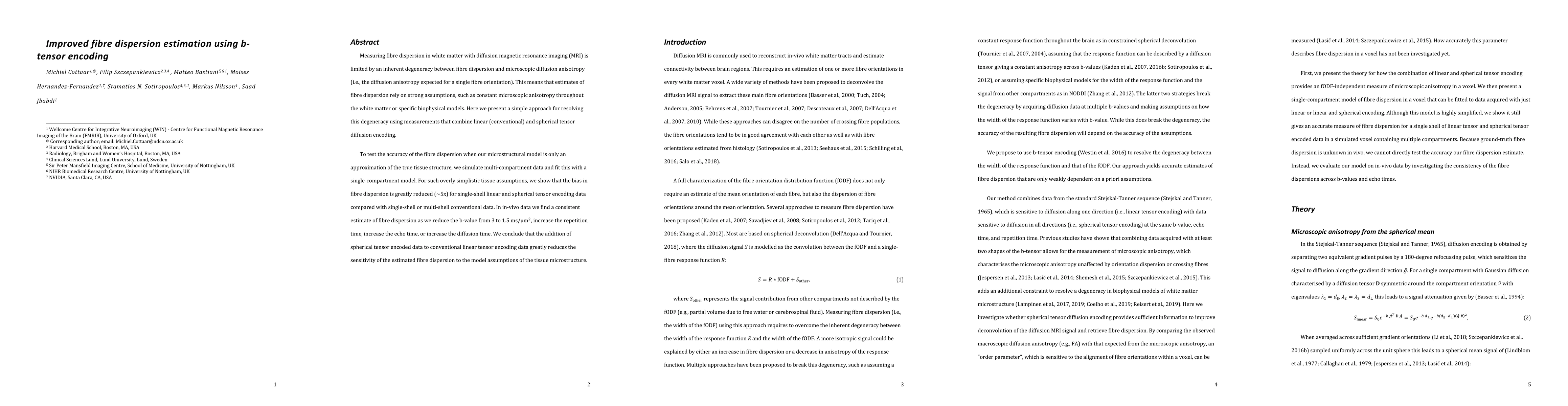
Measuring fibre dispersion in white matter with diffusion magnetic resonance imaging (MRI) is limited by an inherent degeneracy between fibre dispersion and microscopic diffusion anisotropy (i.e., the diffusion anisotropy expected for a single fibre orientation). This means that estimates of fibre dispersion rely on strong assumptions, such as constant microscopic anisotropy throughout the white matter or specific biophysical models. Here we present a simple approach for resolving this degeneracy using measurements that combine linear (conventional) and spherical tensor diffusion encoding. To test the accuracy of the fibre dispersion when our microstructural model is only an approximation of the true tissue structure, we simulate multi-compartment data and fit this with a single-compartment model. For such overly simplistic tissue assumptions, we show that the bias in fibre dispersion is greatly reduced ($\sim$5x) for single-shell linear and spherical tensor encoding data compared with single-shell or multi-shell conventional data. In in-vivo data we find a consistent estimate of fibre dispersion as we reduce the b-value from 3 to 1.5 ms/$\mu$m$^2$, increase the repetition time, increase the echo time, or increase the diffusion time. We conclude that the addition of spherical tensor encoded data to conventional linear tensor encoding data greatly reduces the sensitivity of the estimated fibre dispersion to the model assumptions of the tissue microstructure.

Citation & Reference Network Visualization

This graph shows how this paper is connected to other research. The current paper is shown in dark gray, papers that cite it are in green, and papers it references are in blue. You can:

Current Paper
Papers citing this work (Citations)
Papers cited by this work (References)

• Hover over any node to see the paper title

• Click a node to view that paper's details

• Drag nodes to rearrange the graph

• Use the zoom controls (bottom right) to zoom in/out

Citations and References

Title Authors Year Actions
Towards reliable reconstruction of the mouse brain corticothalamic connectivity using diffusion MRI
Tanzil Mahmud Arefin, C. Lee, Zifei Liang, Harikrishna Rallapalli, Y. Wadghiri, D. Turnbull, Jiangyang Zhang
2023
View Paper
Towards reliable reconstruction of the mouse brain thalamocortical connectivity using diffusion MRI
Tanzil Mahmud Arefin, C. Lee, Zifei Liang, Harikrishna Rallapalli, Y. Wadghiri, D. Turnbull, Jiangyang Zhang
2022
View Paper
Comparing myelin-sensitive magnetic resonance imaging measures and resulting g-ratios in healthy and multiple sclerosis brains
Ronja C. Berg, A. Menegaux, T. Amthor, G. Gilbert, M. Mora, Sarah Schlaeger, Viola Pongratz, M. Lauerer, C. Sorg, M. Doneva, I. Vavasour, M. Mühlau, C. Preibisch
2022
View Paper
Bridging the gap between constrained spherical deconvolution and diffusional variance decomposition via tensor-valued diffusion MRI
Philippe Karan, A. Reymbaut, G. Gilbert, M. Descoteaux
2022
View Paper
Separating Glioma Hyperintensities From White Matter by Diffusion-Weighted Imaging With Spherical Tensor Encoding
J. Brabec, F. Durmo, Filip Szczepankiewicz, P. Brynolfsson, Björn Lampinen, A. Rydelius, Linda Knutsson, C. Westin, P. Sundgren, M. Nilsson
2022
View Paper
Quantification of Tissue Microstructure Using Tensor-Valued Diffusion Encoding: Brain and Body
M. Afzali, Lars Mueller, Filip Szczepankiewicz, Derek K. Jones, J. Schneider
2022
View Paper
What’s new and what’s next in diffusion MRI preprocessing
C. Tax, M. Bastiani, J. Veraart, E. Garyfallidis, M. Irfanoglu
2021
View Paper
In vivo Correlation Tensor MRI reveals microscopic kurtosis in the human brain on a clinical 3T scanner
Lisa Novello, R. Henriques, A. Ianuş, T. Feiweier, N. Shemesh, J. Jovicich
2021
View Paper
Multi-tissue spherical deconvolution of tensor-valued diffusion MRI
B. Jeurissen, Filip Szczepankiewicz
2021
View Paper
Nonparametric D-R 1-R 2 distribution MRI of the living human brain
Jan Martin, A. Reymbaut, M. Schmidt, A. Doerfler, M. Uder, F. Laun, D. Topgaard
2021
View Paper
Enabling constrained spherical deconvolution and diffusional variance decomposition with tensor-valued diffusion MRI
Philippe Karan, A. Reymbaut, G. Gilbert, M. Descoteaux
2021
View Paper
Toward nonparametric diffusion‐T1 characterization of crossing fibers in the human brain
A. Reymbaut, J. Critchley, G. Durighel, T. Sprenger, M. Sughrue, K. Bryskhe, D. Topgaard
2020
View Paper
A Signal Peak Separation Index for Axisymmetric B-Tensor Encoding
Gaëtan Rensonnet, Jonathan Rafael-Patino, B. Macq, J. Thiran, G. Girard, Marco Pizzolato Signal Processing Lab, 'Ecole polytechnique f'ed'erale de Lausanne, Lausanne, Switzerland., Icteam Institute, Universit'e catholique de Louvain, Louvain-la-Neuve, Belgium, Center for Computational Imaging, Radiology Department, C. Vaudois, University of Lausanne, D. O. Mathematics, Computer Science, T. Denmark, Kongens Lyngby, Denmark
2020
View Paper
Gradient waveform design for tensor-valued encoding in diffusion MRI
Filip Szczepankiewicz, C. Westin, M. Nilsson
2020
View Paper
Estimating Brain Connectivity With Diffusion-Weighted Magnetic Resonance Imaging: Promise and Peril.
M. D. Grier, Jan Zimmermann, Sarah R. Heilbronner
2020
View Paper
Magic DIAMOND: Multi-fascicle diffusion compartment imaging with tensor distribution modeling and tensor-valued diffusion encoding
A. Reymbaut, A. Caron, G. Gilbert, F. Szczepankiewicz, M. Nilsson, S. Warfield, M. Descoteaux, B. Scherrer
2020
View Paper
Probing Tissue Microarchitecture of the Baby Brain via Spherical Mean Spectrum Imaging
Khoi Minh Huynh, Tiantian Xu, Ye Wu, Xifeng Wang, Geng Chen, Haiyong Wu, Kim-Han Thung, Weili Lin, D. Shen, P. Yap
2019
View Paper
Using GPUs to accelerate computational diffusion MRI: From microstructure estimation to tractography and connectomes
Moisés Hernández-Fernández, I. Reguly, S. Jbabdi, Mike Giles, Stephen M. Smith, S. Sotiropoulos
2018
View Paper

Comments (0)

This website uses cookies to ensure you get the best experience. Learn more