Quick Summary:

This research uses Generative Adversarial Networks (GANs) to create synthetic brain MR images, enhancing the segmentation of glioma regions by 2D U-Nets. Despite some limitations in gradient representation, the synthetic images improved U-Net performance, suggesting a promising approach to tackle scarcity of medical imaging data for glioma diagnosis.

Summary

Every year thousands of patients are diagnosed with a glioma, a type of malignant brain tumor. Physicians use MR images as a key tool in the diagnosis and treatment of these patients. Neural networks show great potential to aid physicians in the medical image analysis. This study investigates the use of varying amounts of synthetic brain T1-weighted (T1), post-contrast T1-weighted (T1Gd), T2-weighted (T2), and T2 Fluid Attenuated Inversion Recovery (FLAIR) MR images created by a generative adversarial network to overcome the lack of annotated medical image data in training separate 2D U-Nets to segment enhancing tumor, peritumoral edema, and necrosis (non-enhancing tumor core) regions on gliomas. These synthetic MR images were assessed quantitively (SSIM=0.79) and qualitatively by a physician who found that the synthetic images seem stronger for delineation of structural boundaries but struggle more when gradient is significant, (e.g. edema signal in T2 modalities). Multiple 2D U-Nets were trained with original BraTS data and differing subsets of a quarter, half, three-quarters, and all synthetic MR images. There was not an obvious correlation between the improvement of values of the metrics in separate validation dataset for each structure and amount of synthetic data added, there is a strong correlation between the amount of synthetic data added and the number of best overall validation metrics. In summary, this study showed ability to generate high quality synthetic Flair, T2, T1, and T1CE MR images using the GAN. Using the synthetic MR images showed encouraging results to improve the U-Net segmentation performance which has the potential to address the scarcity of readily available medical images.
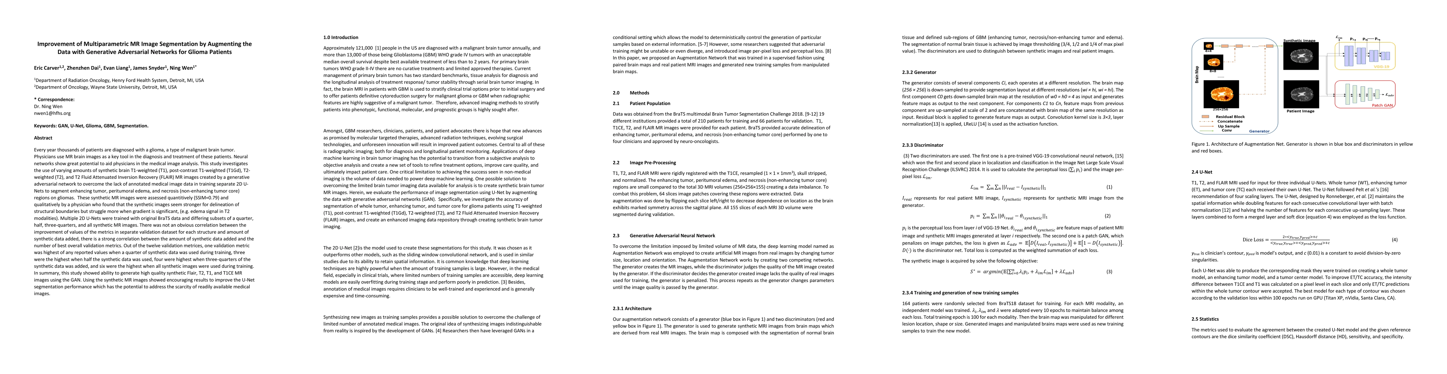
AI Key Findings

Get AI-generated insights about this paper's methodology, results, and significance.

Paper Details

PDF Preview

Download PDF

Key Terms

brain (0.167)
data (0.097)
use (0.081)
study (0.069)
using (0.062)
structure (0.051)
non (0.044)
results (0.036)

Citation Network

Current paper (gray), citations (green), references (blue)

Current Paper
Citations
References
Click to view
Title Authors Year Actions
Generative Adversarial Networks in Brain Imaging: A Decade-Long Review of Progress and Future Directions
Hala Shaari
2025
View Paper
A review of self‐supervised, generative, and few‐shot deep learning methods for data‐limited magnetic resonance imaging segmentation
Zelong Liu, Komal Kainth, Alexander Zhou, T. Deyer, Z. Fayad, H. Greenspan, X. Mei
2024
View Paper
Evaluating synthetic neuroimaging data augmentation for automatic brain tumour segmentation with a deep fully-convolutional network
Fawad Asadi, Thanate Angsuwatanakul, Jamie A O’Reilly
2023
View Paper
The use of generative adversarial networks in medical image augmentation
Ahmed Makhlouf, Marina Maayah, Nada Abughanam, C. Catal
2023
View Paper
Evaluating clinical diversity and plausibility of synthetic capsule endoscopic images
Anuja Vats, Marius Pedersen, A. Mohammed, Ø. Hovde
2023
View Paper
Enhancing cancer differentiation with synthetic MRI examinations via generative models: a systematic review
Avtantil Dimitriadis, Eleftherios Trivizakis, N. Papanikolaou, M. Tsiknakis, K. Marias
2022
View Paper
Deep reinforcement learning and its applications in medical imaging and radiation therapy: a survey
Lanyu Xu, Simeng Zhu, Ning Wen
2022
View Paper
Potential of Deep Learning in Quantitative Magnetic Resonance Imaging for Personalized Radiotherapy.
O. Gurney-Champion, G. Landry, K. Redalen, D. Thorwarth
2022
View Paper
The role of generative adversarial networks in brain MRI: a scoping review
Hazrat Ali, Md. Rafiul Biswas, Farida Mohsen, Uzair Shah, Asma Alamgir, Osama Mousa, Zubair Shah
2022
View Paper
Application of Artificial Intelligence in Diagnosis of Craniopharyngioma
Caijie Qin, Wenxing Hu, Xinsheng Wang, Xibo Ma
2022
View Paper
Magnetic resonance image-based brain tumour segmentation methods: A systematic review
Jayendra M Bhalodiya, S. N. Lim Choi Keung, Theodoros N. Arvanitis
2022
View Paper
Narrative review of generative adversarial networks in medical and molecular imaging
K. Koshino, R. Werner, M. Pomper, R. Bundschuh, F. Toriumi, T. Higuchi, S. Rowe
2021
View Paper
Application of Artificial Intelligence in Diagnosis of Craniopharyngioma
Matteo Zoli, Xinsheng Wang, Xibo Ma, Caijie Qin, Wenxing Hu
View Paper

Comments (0)

This website uses cookies to ensure you get the best experience. Learn more