Quick Summary:

A new method is proposed to improve self-supervised blind spot denoising for microscopy images by incorporating light microscopy's diffraction-limits into the neural network, eliminating high-frequency artifacts and achieving results comparable to supervised methods.

Summary

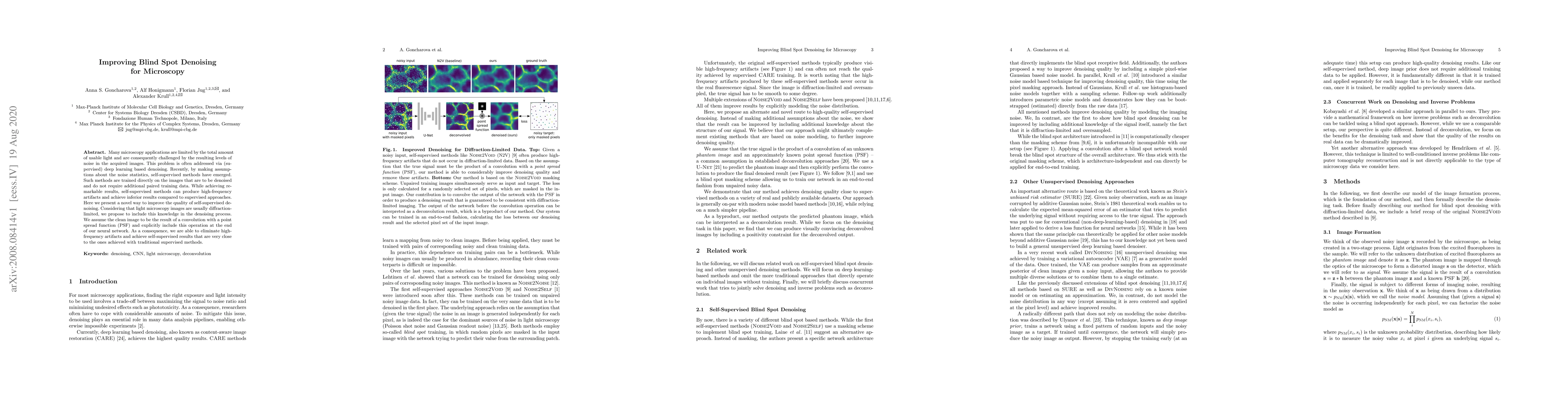
Many microscopy applications are limited by the total amount of usable light and are consequently challenged by the resulting levels of noise in the acquired images. This problem is often addressed via (supervised) deep learning based denoising. Recently, by making assumptions about the noise statistics, self-supervised methods have emerged. Such methods are trained directly on the images that are to be denoised and do not require additional paired training data. While achieving remarkable results, self-supervised methods can produce high-frequency artifacts and achieve inferior results compared to supervised approaches. Here we present a novel way to improve the quality of self-supervised denoising. Considering that light microscopy images are usually diffraction-limited, we propose to include this knowledge in the denoising process. We assume the clean image to be the result of a convolution with a point spread function (PSF) and explicitly include this operation at the end of our neural network. As a consequence, we are able to eliminate high-frequency artifacts and achieve self-supervised results that are very close to the ones achieved with traditional supervised methods.

AI Key Findings

Get AI-generated insights about this paper's methodology, results, and significance.

Paper Details

PDF Preview

Download PDF

Key Terms

supervised (0.274)
self supervised (0.273)
denoising (0.254)
artifacts (0.203)
high frequency (0.203)
microscopy (0.181)
self (0.171)
images (0.165)
include (0.146)
limited (0.140)
methods (0.138)
noise (0.133)
achieve (0.131)
frequency (0.130)
light (0.121)
clean (0.121)
results compared (0.117)
convolution (0.116)
eliminate (0.115)
present novel (0.113)

Citation Network

Current paper (gray), citations (green), references (blue)

Current Paper
Citations
References
Click to view
Title Authors Year Actions
Self-Calibrated Variance-Stabilizing Transformations for Real-World Image Denoising
Sébastien Herbreteau, Michael Unser
2024
View Paper
Connecto-informatics at the mesoscale: current advances in image processing and analysis for mapping the brain connectivity
Yoon Kyoung Choi, Linqing Feng, Won-Ki Jeong, Jinhyun Kim
2024
View Paper
Self-Supervised Joint Learning for pCLE Image Denoising
Kun Yang, Haojie Zhang, Yufei Qiu, Tong Zhai, Zhiguo Zhang
2024
View Paper
Cellpose3: one-click image restoration for improved cellular segmentation
C. Stringer, Marius Pachitariu
2024
View Paper
DeepContrast: Deep Tissue Contrast Enhancement using Synthetic Data Degradations and OOD Model Predictions
N. P. Martins, Y. Kalaidzidis, M. Zerial, Florian Jug
2023
View Paper
A Self-Supervised Denoising Method for pCLE Images
Kun Yang, Haojie Zhang, Zhiguo Zhang, Qingge Sang, Tong Zhai, Yufei Qiu
2023
View Paper
M-Denoiser: Unsupervised image denoising for real-world optical and electron microscopy data
Xiaoya Chong, Min Cheng, Wenqi Fan, Qing Li, Howard Leung
2023
View Paper
μSplit: image decomposition for fluorescence microscopy
Ashesh, Alexander Krull, M. D. Sante, F. Pasqualini, Florian Jug
2022
View Paper
Image denoising in the deep learning era
S. Izadi, D. Sutton, G. Hamarneh
2022
View Paper
MYO10-filopodia support basement membranes at pre-invasive tumor boundaries.
E. Peuhu, Guillaume Jacquemet, C. Scheele, A. Isomursu, Marie-Catherine Laisne, Leena M. Koskinen, I. Paatero, K. Thol, M. Georgiadou, Camilo Guzmán, Satu Koskinen, A. Laiho, L. Elo, P. Boström, P. Hartiala, J. van Rheenen, J. Ivaska
2022
View Paper
Deep Deconvolution Applied to Distributed Acoustic Sensing for Traffic Analysis
M. Ende, A. Ferrari, A. Sladen, C. Richard
2022
View Paper
3D Convolutional Neural Network Framework with Deep Learning for Nuclear Medicine
P. Manimegalai, R. Suresh Kumar, Prajoona Valsalan, R. Dhanagopal, P. V. Vasanth Raj, Jerome Christhudass
2022
View Paper
Deep learning – promises for 3D nuclear imaging: a guide for biologists
Guillaume Mougeot, Tristan Dubos, F. Chausse, E. Péry, K. Graumann, C. Tatout, D. Evans, S. Desset
2022
View Paper
Filopodia-mediated basement membrane assembly at pre-invasive tumor boundaries
E. Peuhu, Guillaume Jacquemet, C. Scheele, A. Isomursu, I. Paatero, K. Thol, M. Georgiadou, Camilo Guzmán, Satu Koskinen, A. Laiho, L. Elo, P. Boström, P. Hartiala, J. van Rheenen, J. Ivaska
2022
View Paper
Deep-learning microscopy image reconstruction with quality control reveals second-scale rearrangements in RNA polymerase II clusters
H. Hajiabadi, I. Mamontova, Roshan Prizak, Agnieszka Pancholi, Anne Koziolek, Lennart Hilbert
2021
View Paper
Real-time Image Denoising of Mixed Poisson-Gaussian Noise in Fluorescence Microscopy Images using ImageJ
Varun Mannam, Yide Zhang, Yinhao Zhu, Evan L. Nichols, Qingfei Wang, Vignesh Sundaresan, Siyuan Zhang, Cody J. Smith, P. Bohn, S. Howard
2021
View Paper
Avoiding a replication crisis in deep-learning-based bioimage analysis
Romain F. Laine, Ignacio Arganda-Carreras, Ricardo Henriques, Guillaume Jacquemet
2021
View Paper
Imaging in focus: An introduction to denoising bioimages in the era of deep learning
Romain F. Laine, Guillaume Jacquemet, Alexander Krull
2021
View Paper
Semi- and Self-Supervised Multi-View Fusion of 3D Microscopy Images using Generative Adversarial Networks
Canyu Yang, Dennis Eschweiler, J. Stegmaier
2021
View Paper
Interpretable Unsupervised Diversity Denoising and Artefact Removal
M. Prakash, M. Delbracio, P. Milanfar, Florian Jug
2021
View Paper
Application of Fast Non-Local Means Algorithm for Noise Reduction Using Separable Color Channels in Light Microscopy Images
S. Kang, Ji-Youn Kim
2021
View Paper
Joint self-supervised blind denoising and noise estimation
J. Ollion, Charles Ollion, E. Gassiat, Luc Lehéricy, S. Corff
2021
View Paper
Squid: Simplifying Quantitative Imaging Platform Development and Deployment
Hongquan Li, Deepak Krishnamurthy, Ethan Li, P. Vyas, Nibha Akireddy, Chew Chai, Manu Prakash
2020
View Paper
µ Split: efficient image decomposition for microscopy data Ashesh
Ashesh, Alexander Krull, M. D. Sante, F. Pasqualini, Florian Jug
2022
View Paper
Removing Pixel Noises and Spatial Artifacts with Generative Diversity Denoising Methods
M. Prakash, M. Delbracio, P. Milanfar, Florian Jug
2021
View Paper
University of Birmingham Imaging in focus
Romain F. Laine, Guillaume Jacquemet, Alexander Krull
View Paper

Comments (0)

This website uses cookies to ensure you get the best experience. Learn more