Publication
Metrics
AI Quick Summary
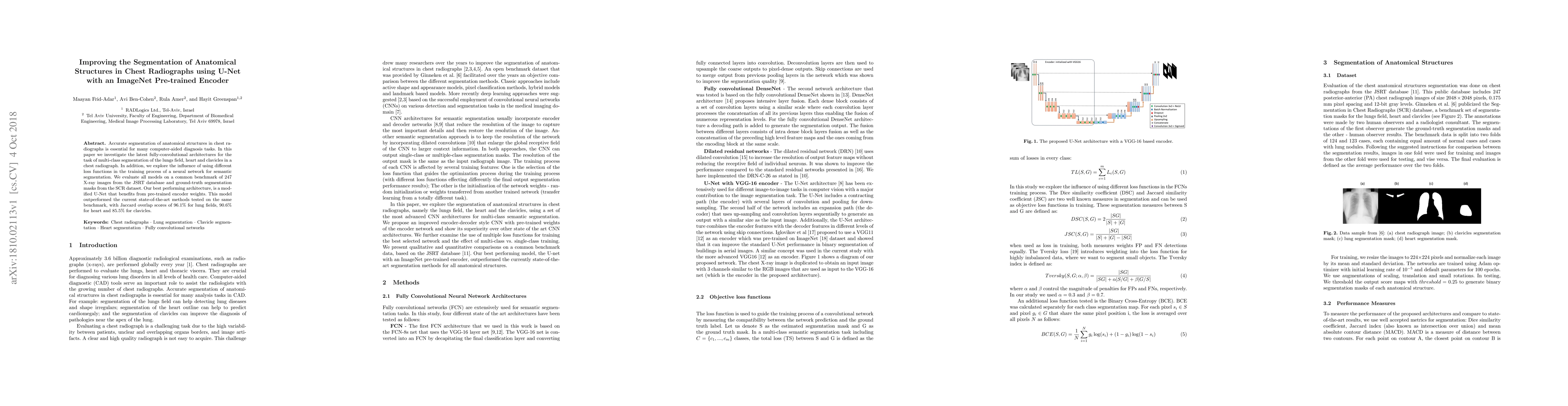
This paper explores the use of a modified U-Net with an ImageNet pre-trained encoder for segmenting anatomical structures in chest radiographs, achieving superior results compared to current methods. The study evaluates different loss functions and reports high Jaccard overlap scores for lung fields, heart, and clavicles.
Paper Preview
Abstract
Accurate segmentation of anatomical structures in chest radiographs is essential for many computer-aided diagnosis tasks. In this paper we investigate the latest fully-convolutional architectures for the task of multi-class segmentation of the lungs field, heart and clavicles in a chest radiograph. In addition, we explore the influence of using different loss functions in the training process of a neural network for semantic segmentation. We evaluate all models on a common benchmark of 247 X-ray images from the JSRT database and ground-truth segmentation masks from the SCR dataset. Our best performing architecture, is a modified U-Net that benefits from pre-trained encoder weights. This model outperformed the current state-of-the-art methods tested on the same benchmark, with Jaccard overlap scores of 96.1% for lung fields, 90.6% for heart and 85.5% for clavicles.
AI Key Findings
Get AI-generated insights about this paper's methodology, results, significance, and more — seven facets brought into focus.
Impact
Paper Details
PDF Preview
Key Terms
Citation Network
Current paper (gray), citations (green), references (blue)
Display is limited for performance on very large graphs.
Discussion 0