Authors
Summary
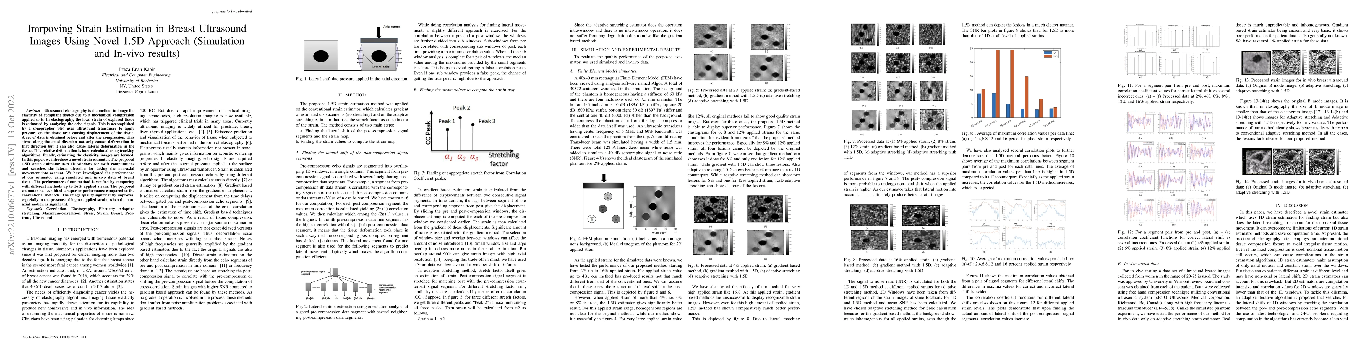
Ultrasound elastography is the method to image the elasticity of compliant tissues due to a mechanical compression applied to it. In elastography, the local strain of explored tissue is estimated by analyzing the echo signals. This is accomplished by a sonographer who uses ultrasound transducer to apply pressure on the tissue area causing displacement of the tissue. A set of data is obtained before and after the compression. This stress along the axial direction not only causes deformation in that direction but it can also cause lateral deformation in the tissue. This relative deformation is later calculated using tracking algorithms. Finally, estimating the elasticity, images are formed. In this paper, we introduce a novel strain estimator. The proposed 1.5D strain estimator uses 1D windows for swift computations and searches the lateral direction for taking the non-axial movement into account. We have investigated the performance of our estimator using simulated and in-vivo data of breast tissue. The performance of our method is verified by comparing with different methods up to 16\% applied strain. The proposed estimator has exhibited a superior performance compared to the conventional methods. The image quality significantly improves, especially in the presence of higher applied strain, when the non-axial motion is significant.
AI Key Findings
Get AI-generated insights about this paper's methodology, results, and significance.
Paper Details
PDF Preview
Key Terms
Citation Network
Current paper (gray), citations (green), references (blue)
Display is limited for performance on very large graphs.
Similar Papers
Found 4 papersAn Adaptive Strain Estimation Algorithm Using Short Term Cross Correlation Kernels and 1.5D Lateral Search
Rasheed Abid, S. Kaisar Alam, Shaiban Ahmed
Investigating Pulse-Echo Sound Speed Estimation in Breast Ultrasound with Deep Learning
Nassir Navab, Vasiliki Sideri-Lampretsa, Magdalini Paschali et al.
| Title | Authors | Year | Actions |
|---|
Comments (0)