Summary
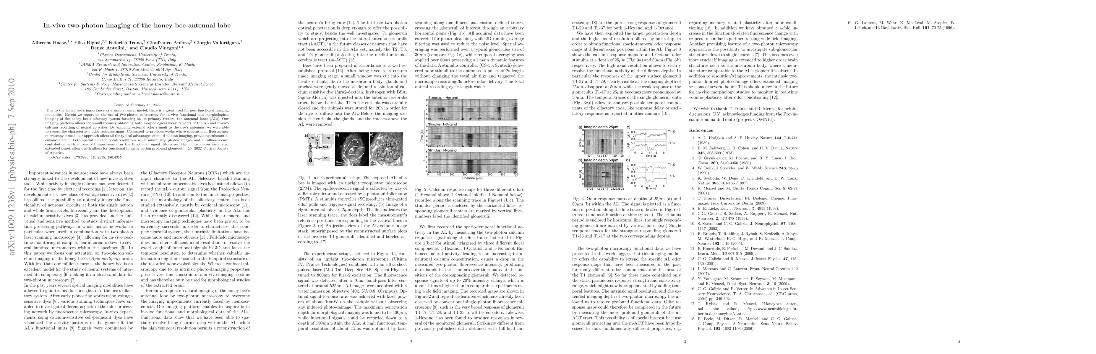
Due to the honey bee's importance as a simple neural model, there is a great need for new functional imaging modalities. Herein we report on the use of two-photon microscopy for in-vivo functional and morphological imaging of the honey bee's olfactory system focusing on its primary centers, the antennal lobes (ALs). Our imaging platform allows for simultaneously obtaining both morphological measurements of the AL and in-vivo calcium recording of neural activities. By applying external odor stimuli to the bee's antennas, we were able to record the characteristic odor response maps. Compared to previous works where conventional fluorescence microscopy is used, our approach offers all the typical advantages of multi-photon imaging, providing substantial enhancement in both spatial and temporal resolutions while minimizing photo-damages and autofluorescence contribution with a four-fold improvement in the functional signal. Moreover, the multi-photon associated extended penetration depth allows for functional imaging within profound glomeruli.
AI Key Findings
Get AI-generated insights about this paper's methodology, results, and significance.
Paper Details
PDF Preview
Key Terms
Citation Network
Current paper (gray), citations (green), references (blue)
Display is limited for performance on very large graphs.
Similar Papers
Found 4 papersImpacts of seasonality and parasitism on honey bee population dynamics
Jun Chen, Jon Harrison, Yun Kang et al.
First large-scale genomic prediction in the honey bee
Manuel Du, Richard Bernstein, Andreas Hoppe et al.
USE OF STINGLESS BEE HONEY (MELIPONINAE SUBFAMILY) IN WOUND TREATMENT: A SYSTEMATIC REVIEW AND META-ANALYSIS
de Medeiros, K. S., Ferreira, F. S., da Silva, D. F. et al.
| Title | Authors | Year | Actions |
|---|
Comments (0)