Publication
Metrics
AI Quick Summary
This paper investigates how varying the laser spot size at the diffuser plane affects the longitudinal spatial coherence function in optical coherence microscopy systems. It finds that an optimal spot size of 3.5 mm yields the highest axial resolution (~13 microns) and suggests that pseudo-thermal light sources can achieve high resolution without requiring complex optical corrections.
Paper Preview
Abstract
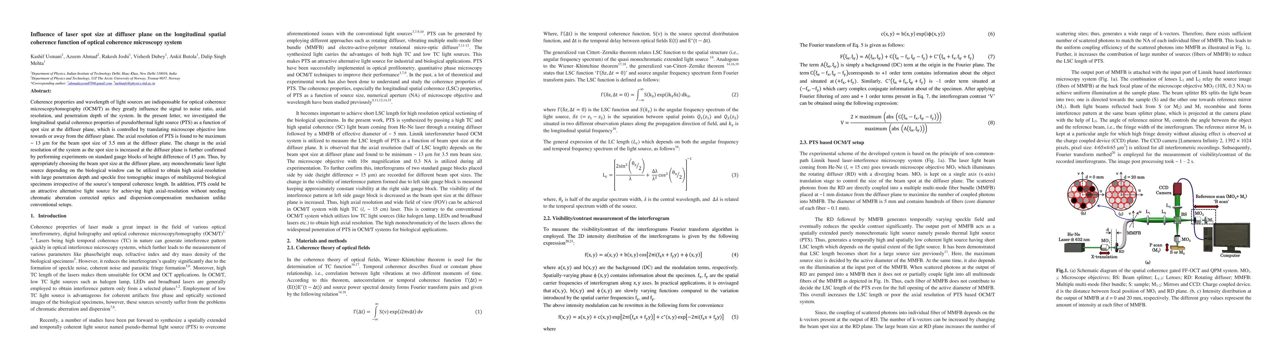
Coherence properties and wavelength of light sources are indispensable for optical coherence microscopy/tomography as they greatly influence the signal to noise ratio, axial resolution, and penetration depth of the system. In the present letter, we investigated the longitudinal spatial coherence properties of the pseudo-thermal light source (PTS) as a function of spot size at the diffuser plane, which is controlled by translating microscope objective lens towards or away from the diffuser plane. The axial resolution of PTS is found to be maximum ~ 13 microns for the beam spot size of 3.5 mm at the diffuser plane. The change in the axial resolution of the system as the spot size is increased at the diffuser plane is further confirmed by performing experiments on standard gauge blocks of height difference of 15 microns. Thus, by appropriately choosing the beam spot size at the diffuser plane, any monochromatic laser light source depending on the biological window can be utilized to obtain high axial-resolution with large penetration depth and speckle-free tomographic images of multilayered biological specimens irrespective of the source temporal coherence length. In addition, PTS could be an attractive alternative light source for achieving high axial-resolution without needing chromatic aberration corrected optics and dispersion-compensation mechanism, unlike conventional setups.
AI Key Findings
Get AI-generated insights about this paper's methodology, results, significance, and more — seven facets brought into focus.
Impact
Paper Details
Authors
PDF Preview
Key Terms
Citation Network
Current paper (gray), citations (green), references (blue)
Display is limited for performance on very large graphs.
Discussion 0