ArxivLens

Quick Summary:

This study assesses initial conditions for reaction-diffusion models predicting glioma growth by comparing MRI-visible abnormalities with histological analysis. Findings suggest that while an exponential decrease in tumor cell density with distance from the core is plausible, edema outlines on MRI do not correspond to cell density iso-contours, and commonly used density values at these outlines are likely overestimated.

DOI: 10.3390/tomography7040055
arXiv Id: 2102.01719
Date Published: 2021-11-23
Date Updated: 2021-11-23

Key Terms (TF-IDF):

tumor (0.326)
mri (0.297)
cell (0.277)
cell (0.270)
visible (0.204)
visible (0.194)
mr (0.192)
diagnosis (0.177)
density (0.174)
brain (0.173)
density (0.173)
maps (0.169)
brain (0.165)
maps (0.165)
core (0.163)
core (0.163)
reaction (0.158)
reaction (0.150)
distance (0.148)
distance (0.147)
License: http://arxiv.org/licenses/nonexclusive-distrib/1.0/

Summary:

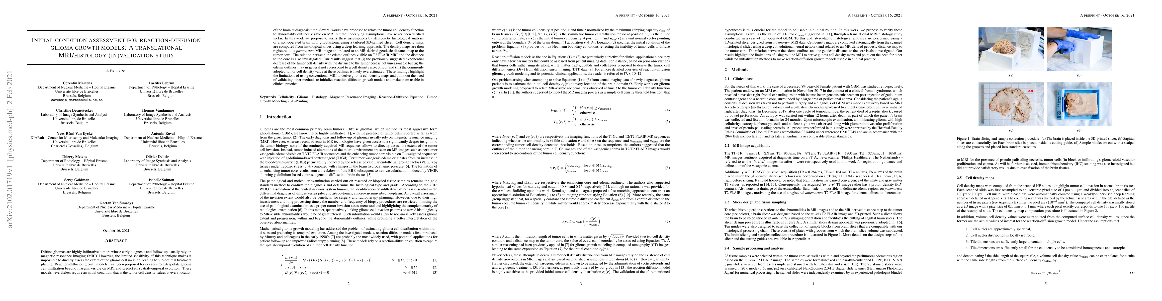
Diffuse gliomas are highly infiltrative tumors whose early diagnosis and follow-up usually rely on magnetic resonance imaging (MRI). However, the limited sensitivity of this technique makes it impossible to directly assess the extent of the glioma cell invasion, leading to sub-optimal treatment planing. Reaction-diffusion growth models have been proposed for decades to extrapolate glioma cell infiltration beyond margins visible on MRI and predict its spatial-temporal evolution. These models nevertheless require an initial condition, that is the tumor cell density values at every location of the brain at diagnosis time. Several works have proposed to relate the tumor cell density function to abnormality outlines visible on MRI but the underlying assumptions have never been verified so far. In this work we propose to verify these assumptions by stereotactic histological analysis of a non-operated brain with glioblastoma using a tailored 3D-printed slicer. Cell density maps are computed from histological slides using a deep learning approach. The density maps are then registered to a postmortem MR image and related to an MR-derived geodesic distance map to the tumor core. The relation between the edema outlines visible on T2 FLAIR MRI and the distance to the core is also investigated. Our results suggest that (i) the previously suggested exponential decrease of the tumor cell density with the distance to the tumor core is not unreasonable but (ii) the edema outlines may in general not correspond to a cell density iso-contour and (iii) the commonly adopted tumor cell density value at these outlines is likely overestimated. These findings highlight the limitations of using conventional MRI to derive glioma cell density maps and point out the need of validating other methods to initialize reaction-diffusion growth models and make them usable in clinical practice.

Citation & Reference Network Visualization

This graph shows how this paper is connected to other research. The current paper is shown in dark gray, papers that cite it are in green, and papers it references are in blue. You can:

Current Paper
Papers citing this work (Citations)
Papers cited by this work (References)

• Hover over any node to see the paper title

• Click a node to view that paper's details

• Drag nodes to rearrange the graph

• Use the zoom controls (bottom right) to zoom in/out

Citations and References

Title Authors Year Actions
Brain tumor detection using proper orthogonal decomposition integrated with deep learning networks
Rita E. Appiah, V. Pulletikurthi, Antonio Esquivel-Puentes, Cristiano Cabrera, N. Hasan, S. Dharmarathne, Luis J. Gomez, Luciano Castillo
2024
View Paper
Toward image-based personalization of glioblastoma therapy: A clinical and biological validation study of a novel, deep learning-driven tumor growth model
M. Metz, Ivan Ezhov, J. Peeken, Josef A. Buchner, Jana Lipková, F. Kofler, Diana Waldmannstetter, C. Delbridge, Christian D. Diehl, Denise Bernhardt, F. Schmidt-Graf, J. Gempt, Stephanie E. Combs, Claus Zimmer, Bjoern H Menze, B. Wiestler
2023
View Paper
Deep Learning for Reaction-Diffusion Glioma Growth Modeling: Towards a Fully Personalized Model?
C. Martens, A. Rovai, Daniele Bonatto, T. Metens, O. Debeir, C. Decaestecker, S. Goldman, G. V. Simaeys
2021
View Paper

Similar Papers

Treatment-aware Diffusion Probabilistic Model for Longitudinal MRI Generation and Diffuse Glioma Growth Prediction

Till Schellhorn, Karoline Skogen, Atle Bjørnerud, Qinghui Liu, Elies Fuster-Garcia, Ivar Thokle Hovden, Donatas Sederevicius, Bradley J MacIntosh, Edvard Grødem, Petter Brandal, Kyrre Eeg Emblem

Published: 2023-09-15

The 2024 Brain Tumor Segmentation (BraTS) Challenge: Glioma Segmentation on Post-treatment MRI

Benedikt Wiestler, Marius George Linguraru, Hongwei Li, Anahita Fathi Kazerooni, Maruf Adewole, Jake Albrecht, Udunna Anazodo, Evan Calabrese, Verena Chung, Keyvan Farahani, Florian Kofler, Dominic LaBella, Bjoern Menze, Ahmed W. Moawad, Jeffrey D. Rudie, Rachit Saluja, Mariam Aboian, Spyridon Bakas, Ayman Nada, Javier Villanueva-Meyer, Jaeyoung Cho, Martin Bendszus, Gianluca Brugnara, Michael Iv, Nico Sollmann, Philipp Vollmuth, Martha Foltyn-Dumitru, Lisa Deptula, Shahriar Faghani, Muhammad Ammar Haider, Talissa Altes, Edvin Isufi, Jaime Gass, Jonathan Thacker, Gian Marco Conte, Mert R. Sabuncu, Pierre Nedelec, John Mongan, Andreas M. Rauschecker, Leo P. Sugrue, Maria Correia de Verdier, Louis Gagnon, Ujjwall Baid, Nourel Hoda Tahon, Jikai Zhang, Maram Alafif, Saif Baig, Ken Chang, Gennaro D'Anna, Diviya Gupta, Ali Hussain, Marinos Kontzialis, Paul Manning, Farzan Moodi, Teresa Nunes, Aaron Simon, David Vu, Rongrong Chai, Eugenio Iglesias, Yury Velichko, Patil Basavasagar, Yaseen Dhemesh, Brandon K. K. Fields, Filip Garrett, Lubomir Hadjiiski, Jona Hattangadi-Gluth, Christopher Hess, Jessica L. Houk, Lester J. Layfield, George Mastorakos, Uyen Nguyen, Sebastian Oliva, Matthew W. Pease, Aditya Rastogi, Jason Sinclair, Robert X. Smith, Igor Vidic, Nathan S. White, Anders Dale, Tyler M. Seibert, Brent Weinberg, Aly Abayazeed, Raymond Huang, Sevcan Turk, Nikdokht Farid

Published: 2024-05-29

Comments (0)

This website uses cookies to ensure you get the best experience. Learn more