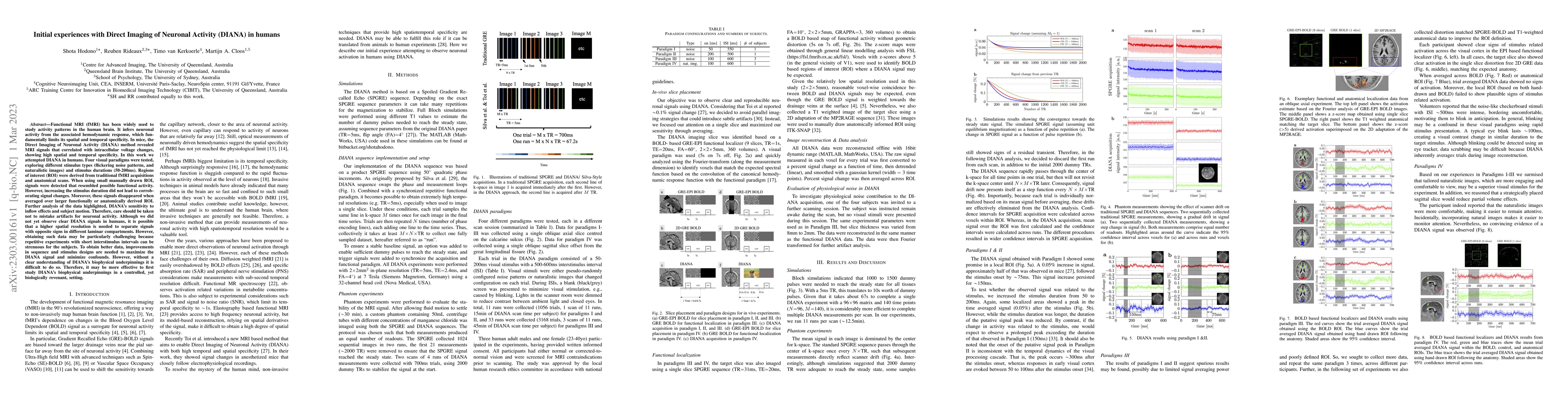
Initial experiences with Direct Imaging of Neuronal Activity (DIANA) in humans
Publication
Metrics
AI Quick Summary
This study attempts Direct Imaging of Neuronal Activity (DIANA) in humans, finding preliminary signals resembling functional activity in small ROIs but not corroborating larger ROIs or longer stimuli. Challenges include sensitivity to artifacts and motion, suggesting a need for higher spatial resolution and improved sequence/stimulus designs.
Paper Preview
Abstract
Functional MRI (fMRI) has been widely used to study activity patterns in the human brain. It infers neuronal activity from the associated hemodynamic response, which fundamentally limits its spatial and temporal specificity. In mice, the Direct Imaging of Neuronal Activity (DIANA) method revealed MRI signals that correlated with intracellular voltage changes, showing high spatial and temporal specificity. In this work we attempted DIANA in humans. Four visual paradigms were tested, exploring different stimulus types (flickering noise patterns, and naturalistic images) and stimulus durations (50-200ms). Regions of interest (ROI) were derived from traditional fMRI acquisitions and anatomical scans. When using small manually drawn ROI, signals were detected that resembled possible functional activity. However, increasing the stimulus duration did not lead to corroborating signal changes. Moreover, these signals disappeared when averaged over larger functionally or anatomically derived ROI. Further analysis of the data highlighted, DIANA's sensitivity to inflow effects and subject motion. Therefore, care should be taken not to mistake artifacts for neuronal activity. Although we did not yet observe clear DIANA signals in humans, it is possible that a higher spatial resolution is needed to separate signals with opposite signs in different laminar compartments. However, obtaining such data may be particularly challenging because repetitive experiments with short interstimulus intervals can be strenuous for the subjects. To obtain better data, improvements in sequence and stimulus designs are needed to maximize the DIANA signal and minimize confounds. However, without a clear understanding of DIANA's biophysical underpinnings it is difficult to do so. Therefore, it may be more effective to first study DIANA's biophysical underpinnings in a controlled, yet biologically revenant, setting.
AI Key Findings
Get AI-generated insights about this paper's methodology, results, significance, and more — seven facets brought into focus.
Impact
Paper Details
Authors
PDF Preview
Key Terms
Citation Network
Current paper (gray), citations (green), references (blue)
Display is limited for performance on very large graphs.
Discussion 0