Integrated Biophysical Modeling and Image Analysis: Application to Neuro-Oncology
Publication
Metrics
AI Quick Summary
This paper explores the integration of biophysical modeling and image analysis to characterize the complex heterogeneity of central nervous system tumors. It combines multi-parametric MRI data with biophysical models to enhance tumor detection, segmentation, and predictive modeling, potentially aiding in future revisions of the WHO classification system for CNS tumors.
Paper Preview
Abstract
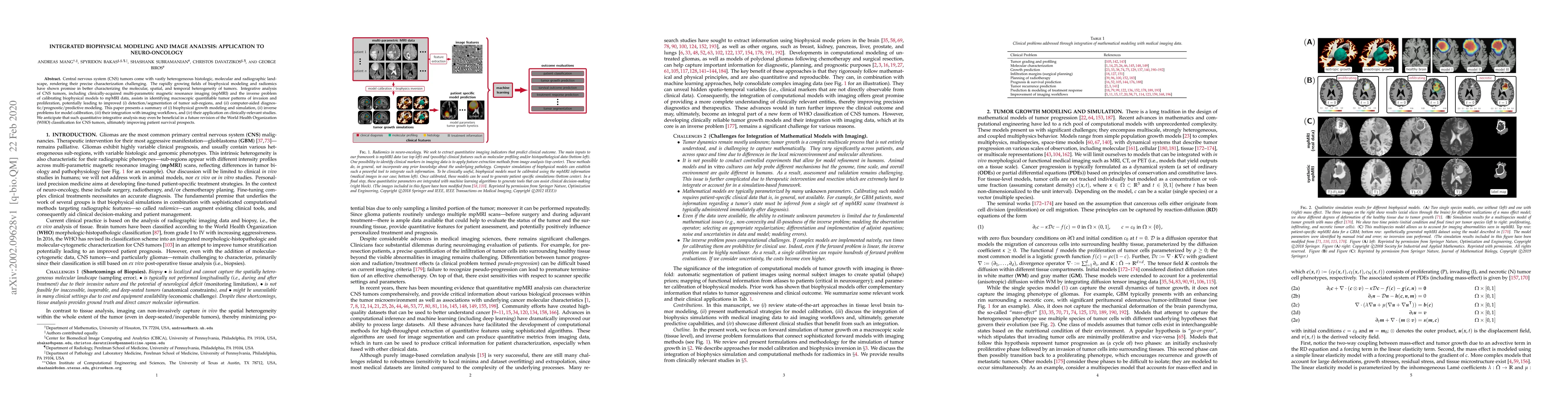
Central nervous system (CNS) tumors come with the vastly heterogeneous histologic, molecular and radiographic landscape, rendering their precise characterization challenging. The rapidly growing fields of biophysical modeling and radiomics have shown promise in better characterizing the molecular, spatial, and temporal heterogeneity of tumors. Integrative analysis of CNS tumors, including clinically-acquired multi-parametric magnetic resonance imaging (mpMRI) and the inverse problem of calibrating biophysical models to mpMRI data, assists in identifying macroscopic quantifiable tumor patterns of invasion and proliferation, potentially leading to improved (i) detection/segmentation of tumor sub-regions, and (ii) computer-aided diagnostic/prognostic/predictive modeling. This paper presents a summary of (i) biophysical growth modeling and simulation, (ii) inverse problems for model calibration, (iii) their integration with imaging workflows, and (iv) their application on clinically-relevant studies. We anticipate that such quantitative integrative analysis may even be beneficial in a future revision of the World Health Organization (WHO) classification for CNS tumors, ultimately improving patient survival prospects.
AI Key Findings
Get AI-generated insights about this paper's methodology, results, significance, and more — seven facets brought into focus.
Impact
Paper Details
Authors
PDF Preview
Key Terms
Citation Network
Current paper (gray), citations (green), references (blue)
Display is limited for performance on very large graphs.
Discussion 0