The importance of rapid and accurate histologic analysis of surgical tissue
in the operating room has been recognized for over a century. Our
standard-of-care intraoperative pathology workflow is based on light microscopy
and H\&E histology, which is slow, resource-intensive, and lacks real-time
digital imaging capabilities. Here, we present an emerging and innovative
method for intraoperative histologic analysis, called Intelligent Histology,
that integrates artificial intelligence (AI) with stimulated Raman histology
(SRH). SRH is a rapid, label-free, digital imaging method for real-time
microscopic tumor tissue analysis. SRH generates high-resolution digital images
of surgical specimens within seconds, enabling AI-driven tumor histologic
analysis, molecular classification, and tumor infiltration detection. We review
the scientific background, clinical translation, and future applications of
intelligent histology in tumor neurosurgery. We focus on the major scientific
and clinical studies that have demonstrated the transformative potential of
intelligent histology across multiple neurosurgical specialties, including
neurosurgical oncology, skull base, spine oncology, pediatric tumors, and
periperal nerve tumors. Future directions include the development of AI
foundation models through multi-institutional datasets, incorporating clinical
and radiologic data for multimodal learning, and predicting patient outcomes.
Intelligent histology represents a transformative intraoperative workflow that
can reinvent real-time tumor analysis for 21st century neurosurgery.
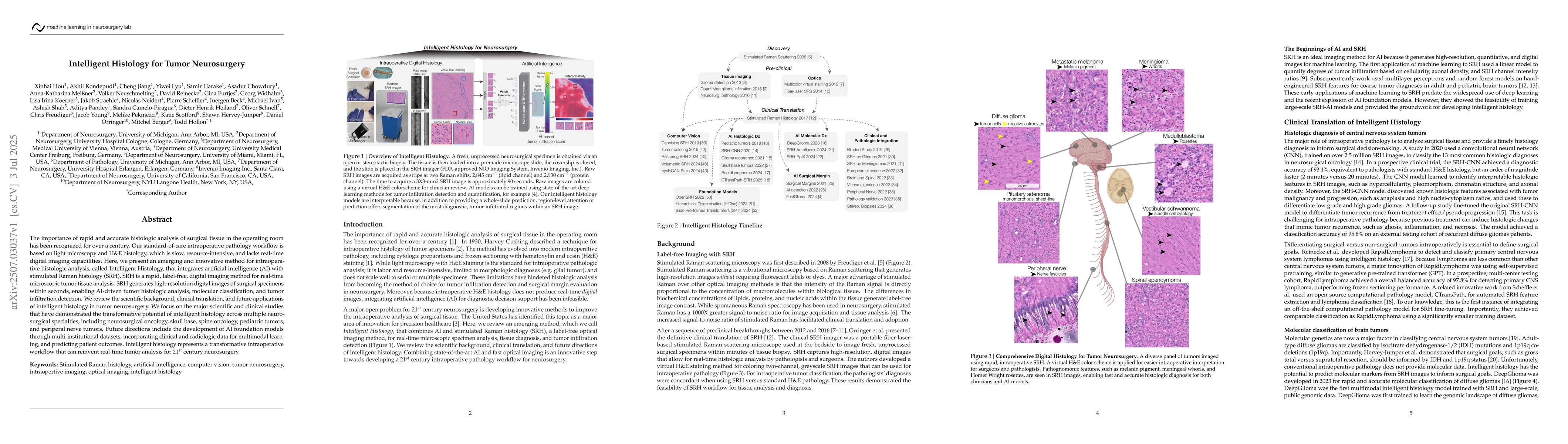
Discussion 0