Interactive Image Selection and Training for Brain Tumor Segmentation Network
Publication
Metrics
AI Quick Summary
This research introduces an interactive image selection and training method based on Feature Learning from Image Markers (FLIM) for brain tumor segmentation. The methodology leverages expert input to train smaller networks effectively, achieving comparable or superior performance to traditional backpropagation methods using fewer training images.
Paper Preview
Abstract
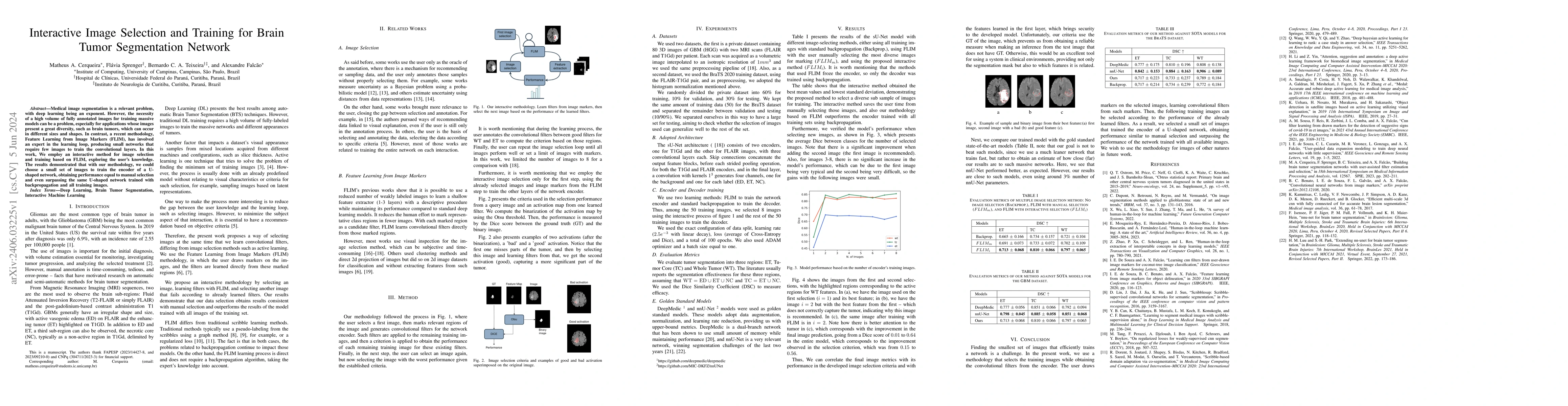
Medical image segmentation is a relevant problem, with deep learning being an exponent. However, the necessity of a high volume of fully annotated images for training massive models can be a problem, especially for applications whose images present a great diversity, such as brain tumors, which can occur in different sizes and shapes. In contrast, a recent methodology, Feature Learning from Image Markers (FLIM), has involved an expert in the learning loop, producing small networks that require few images to train the convolutional layers. In this work, We employ an interactive method for image selection and training based on FLIM, exploring the user's knowledge. The results demonstrated that with our methodology, we could choose a small set of images to train the encoder of a U-shaped network, obtaining performance equal to manual selection and even surpassing the same U-shaped network trained with backpropagation and all training images.
AI Key Findings
Get AI-generated insights about this paper's methodology, results, significance, and more — seven facets brought into focus.
Impact
Paper Details
Authors
PDF Preview
Key Terms
Citation Network
Current paper (gray), citations (green), references (blue)
Display is limited for performance on very large graphs.
Discussion 0