Interactive Medical Image Segmentation via Point-Based Interaction and Sequential Patch Learning
Publication
Metrics
AI Quick Summary
This paper introduces an innovative interactive medical image segmentation method that uses point-based interaction and sequential patch learning. It employs a ConvRNN network with a gated memory unit to learn bi-directional sequential patches, requiring only a central point click for initialization, thus enhancing performance and reducing segmentation time.
Paper Preview
Abstract
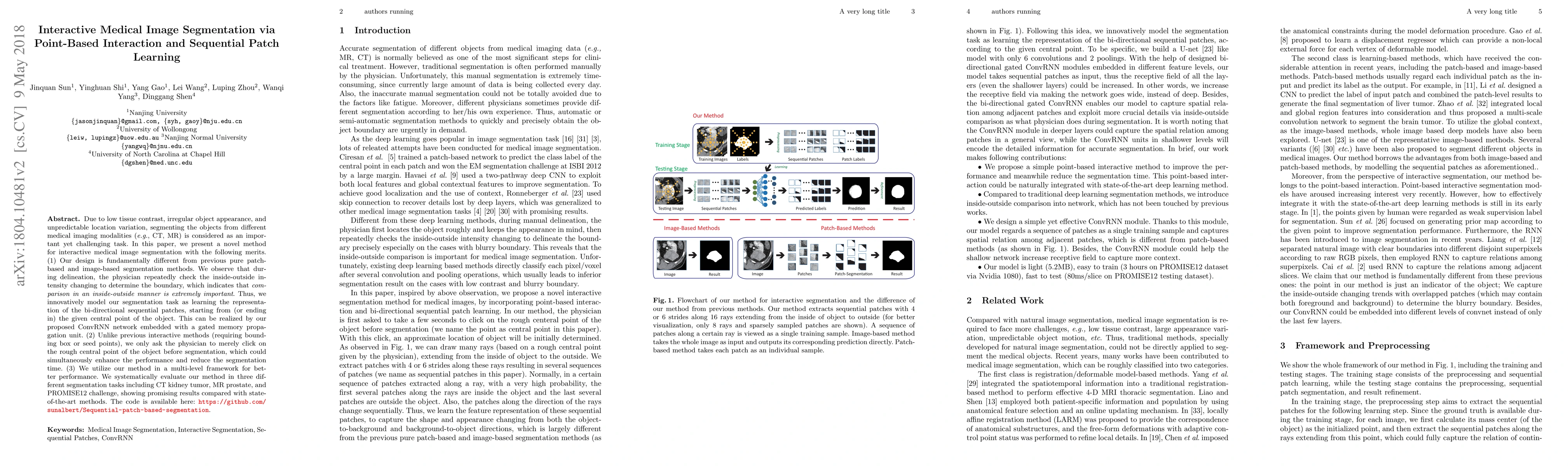
Due to low tissue contrast, irregular object appearance, and unpredictable location variation, segmenting the objects from different medical imaging modalities (e.g., CT, MR) is considered as an important yet challenging task. In this paper, we present a novel method for interactive medical image segmentation with the following merits. (1) Our design is fundamentally different from previous pure patch-based and image-based segmentation methods. We observe that during delineation, the physician repeatedly check the inside-outside intensity changing to determine the boundary, which indicates that comparison in an inside-outside manner is extremely important. Thus, we innovatively model our segmentation task as learning the representation of the bi-directional sequential patches, starting from (or ending in) the given central point of the object. This can be realized by our proposed ConvRNN network embedded with a gated memory propagation unit. (2) Unlike previous interactive methods (requiring bounding box or seed points), we only ask the physician to merely click on the rough central point of the object before segmentation, which could simultaneously enhance the performance and reduce the segmentation time. (3) We utilize our method in a multi-level framework for better performance. We systematically evaluate our method in three different segmentation tasks including CT kidney tumor, MR prostate, and PROMISE12 challenge, showing promising results compared with state-of-the-art methods. The code is available here: \href{https://github.com/sunalbert/Sequential-patch-based-segmentation}{Sequential-patch-based-segmentation}.
AI Key Findings
Get AI-generated insights about this paper's methodology, results, significance, and more — seven facets brought into focus.
Impact
Paper Details
PDF Preview
Key Terms
Citation Network
Current paper (gray), citations (green), references (blue)
Display is limited for performance on very large graphs.
Discussion 0