01
MethodologyHow they did it
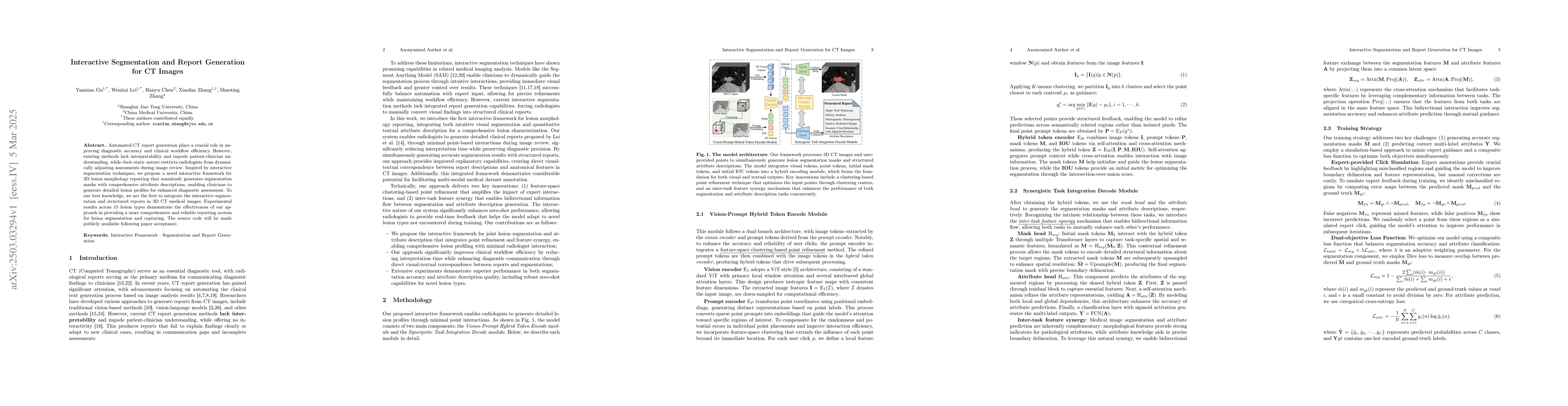
The paper proposes an interactive framework for lesion morphology reporting in CT images, integrating visual segmentation with structured attribute description. It utilizes K-means clustering, a hybrid token encoder, and a decode module for segmentation and attribute prediction. The approach leverages bidirectional feature synergy between tasks and employs an expert-mimicking click simulation for training.
Discussion 0