This study reveals that direct physical contact between fibroblasts and myofibroblasts can drive fibroblast-to-myofibroblast transitions without biochemical or mechanical cues, highlighting a novel mechanism for fibrosis persistence. The inhibition of G{\alpha}q/11/14 with FR9003591 blocks this contact-based transition, suggesting a potential therapeutic approach for fibrosis.
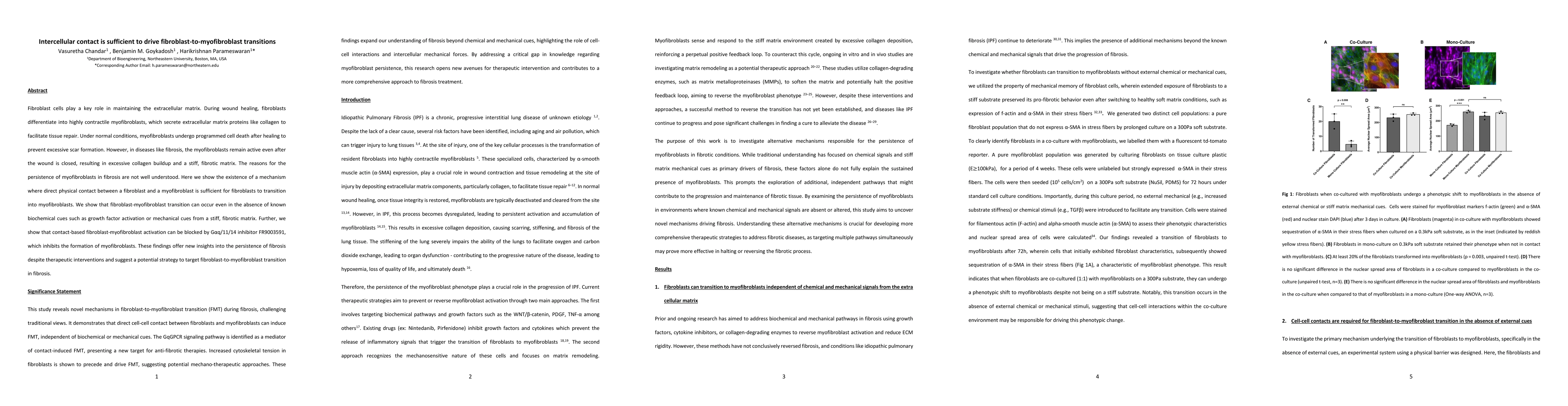
Fibroblast cells play a key role in maintaining the extracellular matrix. During wound healing, fibroblasts differentiate into highly contractile myofibroblasts, which secrete extracellular matrix proteins like collagen to facilitate tissue repair. Under normal conditions, myofibroblasts undergo programmed cell death after healing to prevent excessive scar formation. However, in diseases like fibrosis, the myofibroblasts remain active even after the wound is closed, resulting in excessive collagen buildup and a stiff, fibrotic matrix. The reasons for the persistence of myofibroblasts in fibrosis are not well understood. Here we show the existence of a mechanism where direct physical contact between a fibroblast and a myofibroblast is sufficient for fibroblasts to transition into myofibroblasts. We show that fibroblast-myofibroblast transition can occur even in the absence of known biochemical cues such as growth factor activation or mechanical cues from a stiff, fibrotic matrix. Further, we show that contact-based fibroblast-myofibroblast activation can be blocked by G{\alpha}q/11/14 inhibitor FR9003591, which inhibits the formation of myofibroblasts. These findings offer new insights into the persistence of fibrosis despite therapeutic interventions and suggest a potential strategy to target fibroblast-to-myofibroblast transition in fibrosis.
Generated Jun 10, 2025
The study utilized optically clear PDMS substrates with tunable Young's modulus, NIH 3T3 mouse fibroblasts, co-culture platforms, lentiviral vector encoding tdTomato for fibroblast identification, immunostaining, microscopy, point displacement on the matrix, fluorescent PIP2 imaging, patterning cell ensembles, and traction force microscopy/monolayer stress microscopy measurements.
This research uncovers novel mechanisms driving FMT in fibrosis, challenging traditional views and emphasizing the critical role of intercellular mechanical signaling and the GqGPCR pathway as mediators of contact-induced FMT, presenting a promising target for anti-fibrotic therapies.
The study demonstrates the use of advanced techniques such as optically clear PDMS substrates, fluorescent PIP2 imaging, and traction force/monolayer stress microscopy to investigate cell-cell interactions and mechanical signaling in FMT.
This work reveals a previously unrecognized mechanism of FMT driven by direct cell-cell interactions and mechanical signaling, expanding our understanding of fibrosis progression and opening new avenues for therapeutic interventions targeting the mechanical aspects of fibrotic disease.
Current paper (gray), citations (green), references (blue)
Display is limited for performance on very large graphs.
Pratyush Tiwary, Ziyue Zou, Shams Mehdi et al.
Aguado, Brian A, Vogt, Brandon J, Wang, Peter et al.
No citations found for this paper.
Comments (0)