Intermediate Domain-guided Adaptation for Unsupervised Chorioallantoic Membrane Vessel Segmentation

Source: ArXiv

AI Quick Summary

This paper proposes an Intermediate Domain-guided Adaptation (IDA) method for unsupervised segmentation of vessels in the chorioallantoic membrane (CAM) using retinal images for training. The method employs Multi-Resolution Asymmetric Translation and Intermediate Domain-guided Contrastive Learning to enhance domain adaptation, creating the first CAM dataset for validation and demonstrating superior performance over existing approaches.

Share: Share on X LinkedIn

Summary

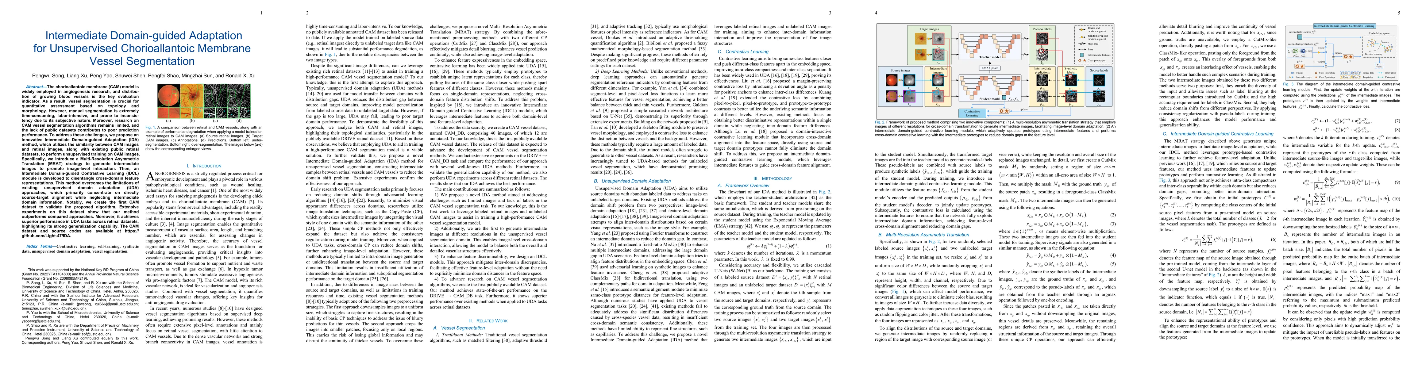
The chorioallantoic membrane (CAM) model is widely employed in angiogenesis research, and distribution of growing blood vessels is the key evaluation indicator. As a result, vessel segmentation is crucial for quantitative assessment based on topology and morphology. However, manual segmentation is extremely time-consuming, labor-intensive, and prone to inconsistency due to its subjective nature. Moreover, research on CAM vessel segmentation algorithms remains limited, and the lack of public datasets contributes to poor prediction performance. To address these challenges, we propose an innovative Intermediate Domain-guided Adaptation (IDA) method, which utilizes the similarity between CAM images and retinal images, along with existing public retinal datasets, to perform unsupervised training on CAM images. Specifically, we introduce a Multi-Resolution Asymmetric Translation (MRAT) strategy to generate intermediate images to promote image-level interaction. Then, an Intermediate Domain-guided Contrastive Learning (IDCL) module is developed to disentangle cross-domain feature representations. This method overcomes the limitations of existing unsupervised domain adaptation (UDA) approaches, which primarily concentrate on directly source-target alignment while neglecting intermediate domain information. Notably, we create the first CAM dataset to validate the proposed algorithm. Extensive experiments on this dataset show that our method outperforms compared approaches. Moreover, it achieves superior performance in UDA tasks across retinal datasets, highlighting its strong generalization capability. The CAM dataset and source codes are available at https://github.com/Light-47/IDA.

AI Key Findings

Generated Jun 10, 2025

Methodology

The research proposes an Intermediate Domain-guided Adaptation (IDA) method for unsupervised chorioallantoic membrane (CAM) vessel segmentation, leveraging similarity with retinal images and public retinal datasets, employing Multi-Resolution Asymmetric Translation (MRAT) for intermediate image generation and Intermediate Domain-guided Contrastive Learning (IDCL) for feature representation disentanglement.

Key Results

  • IDA outperforms existing unsupervised domain adaptation (UDA) methods in CAM vessel segmentation tasks.
  • IDA achieves superior performance in UDA tasks across retinal datasets, demonstrating strong generalization capability.
  • The method introduces the first CAM dataset for validation, contributing to future research in this domain.

Significance

This research is significant as it addresses the challenges of manual, time-consuming, and inconsistent CAM vessel segmentation, crucial for angiogenesis research, by proposing an efficient unsupervised segmentation method that can generalize well across different datasets.

Technical Contribution

The paper presents an innovative IDA framework that combines MRAT for generating intermediate images and IDCL for contrastive learning, effectively reducing domain gaps at both image and feature levels.

Novelty

The novelty of this work lies in its approach to utilizing intermediate domain information for contrastive learning, which enhances cross-domain feature interaction and improves segmentation accuracy in the CAM context.

Limitations

  • The study is limited to UDA tasks and does not explore supervised or semi-supervised approaches.
  • Performance in low-contrast regions, such as certain areas in CHASEDB1 and STARE, still requires refinement.

Future Work

  • Explore the application of the model in angiogenesis-related research for rapid quantitative parameter characterization.
  • Investigate the effectiveness of the proposed method on other medical imaging segmentation tasks.

Paper Details

PDF Preview

Download PDF

Citation Network

Current paper (gray), citations (green), references (blue)

Display is limited for performance on very large graphs.

Current Paper
Citations
References
Click to view

No citations found for this paper.

Comments (0)

This website uses cookies to ensure you get the best experience. Learn more