Publication
Metrics
AI Quick Summary
The paper introduces AEMS-Net, a deep-learning framework for simultaneous prediction of multiple subcellular structures in fluorescence microscopy, reducing imaging delays and improving quality. The model uses interpretable functions and attention mechanisms to enhance real-time live-cell imaging of mitochondria and microtubules.
Paper Preview
Abstract
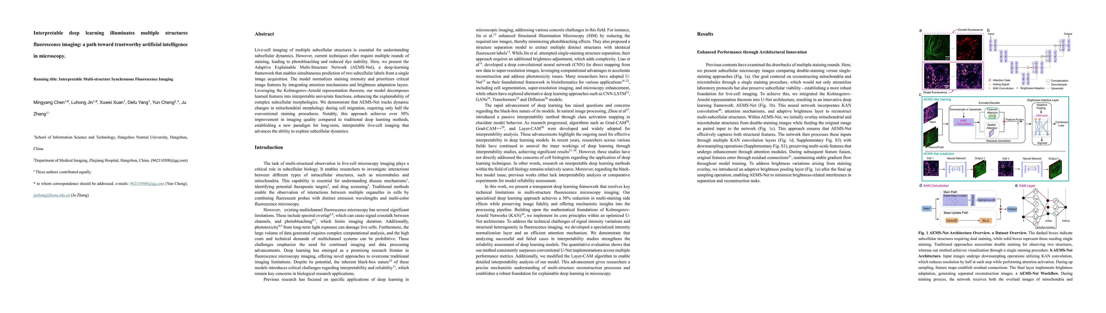
Live-cell imaging of multiple subcellular structures is essential for understanding subcellular dynamics. However, the conventional multi-color sequential fluorescence microscopy suffers from significant imaging delays and limited number of subcellular structure separate labeling, resulting in substantial limitations for real-time live-cell research applications. Here, we present the Adaptive Explainable Multi-Structure Network (AEMS-Net), a deep-learning framework that enables simultaneous prediction of two subcellular structures from a single image. The model normalizes staining intensity and prioritizes critical image features by integrating attention mechanisms and brightness adaptation layers. Leveraging the Kolmogorov-Arnold representation theorem, our model decomposes learned features into interpretable univariate functions, enhancing the explainability of complex subcellular morphologies. We demonstrate that AEMS-Net allows real-time recording of interactions between mitochondria and microtubules, requiring only half the conventional sequential-channel imaging procedures. Notably, this approach achieves over 30% improvement in imaging quality compared to traditional deep learning methods, establishing a new paradigm for long-term, interpretable live-cell imaging that advances the ability to explore subcellular dynamics.
AI Key Findings
Get AI-generated insights about this paper's methodology, results, significance, and more — seven facets brought into focus.
Authors
PDF Preview
Related Papers
No references found for this paper.
Discussion 0