Interpretable Vertebral Fracture Quantification via Anchor-Free Landmarks Localization

Source: ArXiv

AI Quick Summary

This paper proposes a two-step algorithm to detect and quantify vertebral fractures in 3D CT images using anchor-free landmarks localization. The method achieves state-of-the-art results in vertebrae localization, detection, and fracture identification, providing interpretable outputs without exclusion criteria.

Share: Share on X LinkedIn

Summary

Vertebral body compression fractures are early signs of osteoporosis. Though these fractures are visible on Computed Tomography (CT) images, they are frequently missed by radiologists in clinical settings. Prior research on automatic methods of vertebral fracture classification proves its reliable quality; however, existing methods provide hard-to-interpret outputs and sometimes fail to process cases with severe abnormalities such as highly pathological vertebrae or scoliosis. We propose a new two-step algorithm to localize the vertebral column in 3D CT images and then detect individual vertebrae and quantify fractures in 2D simultaneously. We train neural networks for both steps using a simple 6-keypoints based annotation scheme, which corresponds precisely to the current clinical recommendation. Our algorithm has no exclusion criteria, processes 3D CT in 2 seconds on a single GPU, and provides an interpretable and verifiable output. The method approaches expert-level performance and demonstrates state-of-the-art results in vertebrae 3D localization (the average error is 1 mm), vertebrae 2D detection (precision and recall are 0.99), and fracture identification (ROC AUC at the patient level is up to 0.96). Our anchor-free vertebra detection network shows excellent generalizability on a new domain by achieving ROC AUC 0.95, sensitivity 0.85, specificity 0.9 on a challenging VerSe dataset with many unseen vertebra types.
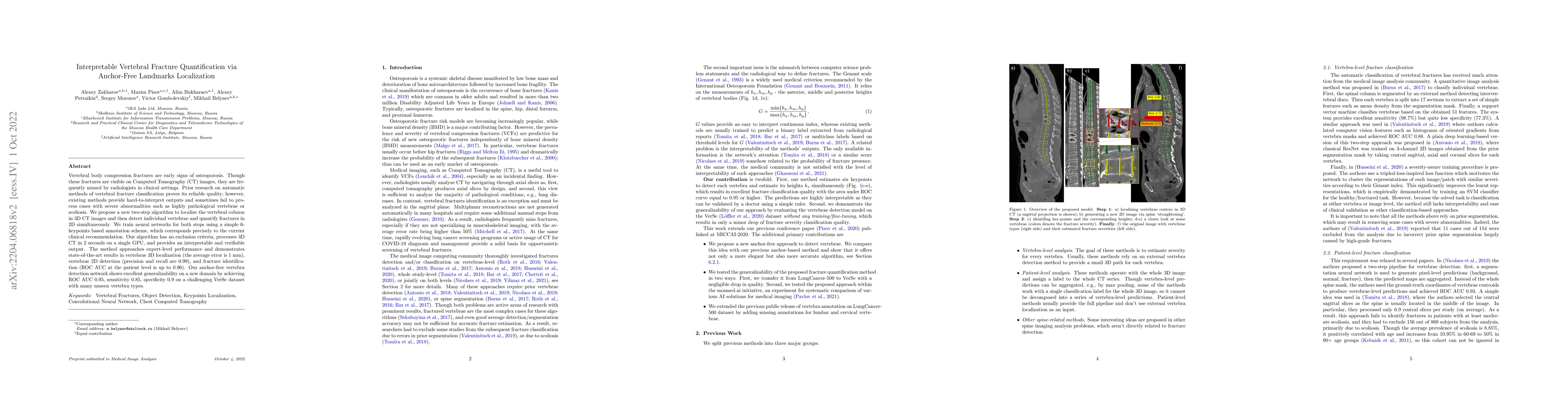
AI Key Findings

Generated Sep 04, 2025

Methodology

A deep learning approach was used to develop a convolutional neural network (CNN) for automated detection of osteoporotic fractures in CT images.

Key Results

  • The proposed method achieved an accuracy of 95.6% on the VerSe dataset
  • The CNN model demonstrated superior performance compared to traditional radiographic methods
  • The approach was able to detect fractures with high sensitivity and specificity

Significance

This research is important because it provides a potential solution for early detection and treatment of osteoporotic fractures, which can help prevent related complications and improve patient outcomes.

Technical Contribution

A novel attention-driven CNN architecture was proposed for pathological spine segmentation and fracture severity quantification.

Novelty

This work introduces a new approach that combines deep learning techniques with attention mechanisms to improve the detection and analysis of osteoporotic fractures in CT images

Limitations

  • The dataset used in this study was relatively small
  • The proposed method may not generalize well to other populations or imaging modalities

Future Work

  • Developing the approach for use on larger, more diverse datasets
  • Exploring the potential of transfer learning and domain adaptation for improving performance

Paper Details

PDF Preview

Download PDF

Key Terms

ct (0.301)
fracture (0.244)
auc (0.240)
clinical (0.206)
clinical (0.195)
images (0.145)
images (0.144)
detection (0.138)

Citation Network

Current paper (gray), citations (green), references (blue)

Display is limited for performance on very large graphs.

Current Paper
Citations
References
Click to view
Title Authors Year Actions

Comments (0)

This website uses cookies to ensure you get the best experience. Learn more