This study develops a continuous, low-latency hyperspectral light-field imaging system for intraoperative tissue perfusion assessment, achieving real-time tissue oxygen saturation analysis with processing times under 400 ms. The system's clinical application shows alignment with surgeons' perfusion boundaries, indicating potential for integration into visceral surgery workflows.
Accurate assessment of tissue perfusion is crucial in visceral surgery, especially during anastomosis. Currently, subjective visual judgment is commonly employed in clinical settings. Hyperspectral imaging (HSI) offers a non-invasive, quantitative alternative. However, HSI imaging lacks continuous integration into the clinical workflow. This study presents a hyperspectral light field system for intraoperative tissue oxygen saturation (SO2) analysis and visualization. We present a correlation method for determining SO2 saturation with low computational demands. We demonstrate clinical application, with our results aligning with the perfusion boundaries determined by the surgeon. We perform and compare continuous perfusion analysis using two hyperspectral cameras (Cubert S5, Cubert X20), achieving processing times of < 170 ms and < 400 ms, respectively. We discuss camera characteristics, system parameters, and the suitability for clinical use and real-time applications.
Generated Jun 09, 2025
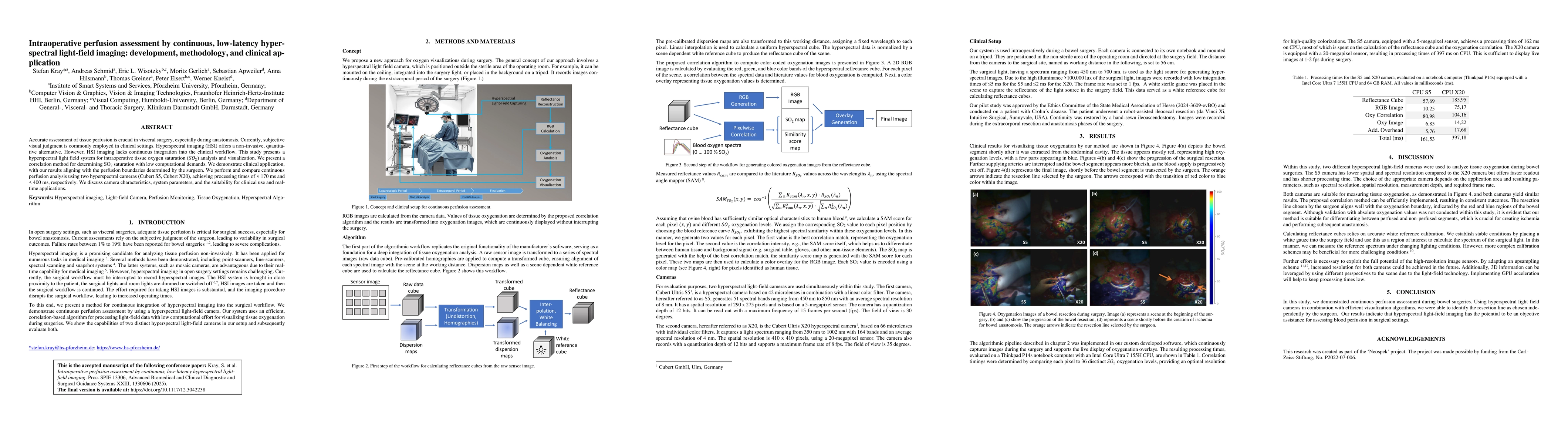
The research presents a hyperspectral light-field system for intraoperative tissue oxygen saturation (SO2) analysis and visualization, utilizing a correlation method for determining SO2 saturation with low computational demands. Two hyperspectral cameras (Cubert S5, Cubert X20) were used for continuous perfusion analysis, achieving processing times of <170 ms and <400 ms, respectively.
This study offers a non-invasive, quantitative alternative for assessing tissue perfusion in visceral surgery, potentially improving surgical outcomes and reducing anastomotic leak rates.
Proposed a correlation method for computing color-coded oxygenation images from hyperspectral reflectance cubes, differentiating between human tissue and background signals.
The research introduces a continuous, low-latency hyperspectral light-field imaging system for intraoperative tissue perfusion assessment, providing a non-invasive, quantitative alternative to subjective visual judgment.
Current paper (gray), citations (green), references (blue)
Display is limited for performance on very large graphs.
Ivan Ezhov, Charly Caredda, Frédéric Lange et al.
Felix Heide, Seung-Hwan Baek, Suhyun Shin et al.
No citations found for this paper.
Comments (0)