Publication
Metrics
AI Quick Summary
This paper presents a method for isotope analysis using a scanning transmission electron microscope, differentiating isotopes of the same element based on the probability of imaging electrons ejecting atoms. The technique is demonstrated in graphene samples and shows a spatial resolution better than 20% in mapping isotope concentration.
Paper Preview
Abstract
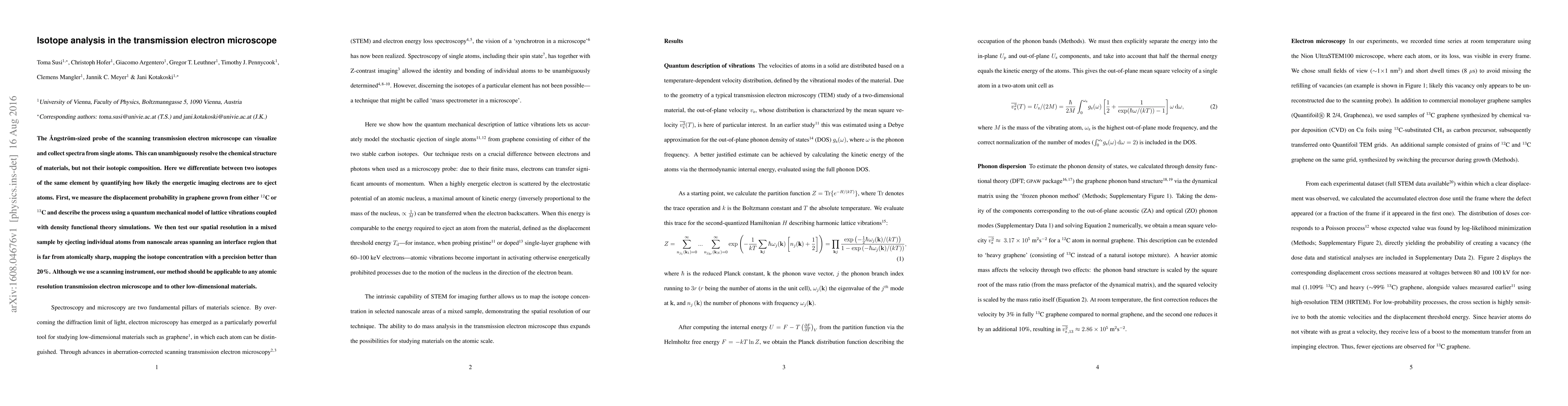
The {\AA}ngstr\"om-sized probe of the scanning transmission electron microscope can visualize and collect spectra from single atoms. This can unambiguously resolve the chemical structure of materials, but not their isotopic composition. Here we differentiate between two isotopes of the same element by quantifying how likely the energetic imaging electrons are to eject atoms. First, we measure the displacement probability in graphene grown from either $^{12}$C or $^{13}$C and describe the process using a quantum mechanical model of lattice vibrations coupled with density functional theory simulations. We then test our spatial resolution in a mixed sample by ejecting individual atoms from nanoscale areas spanning an interface region that is far from atomically sharp, mapping the isotope concentration with a precision better than 20%. Although we use a scanning instrument, our method should be applicable to any atomic resolution transmission electron microscope and to other low-dimensional materials.
AI Key Findings
Get AI-generated insights about this paper's methodology, results, significance, and more — seven facets brought into focus.
Impact
Paper Details
PDF Preview
Key Terms
Citation Network
Current paper (gray), citations (green), references (blue)
Display is limited for performance on very large graphs.
Discussion 0