Joint multi-contrast Variational Network reconstruction (jVN) with application to rapid 2D and 3D imaging
Publication
Metrics
AI Quick Summary
This study introduces a joint multi-contrast variational network (jVN) for reconstructing high-quality MRI images from accelerated multi-channel data, achieving up to 16-fold acceleration with minimal image blurring. The method leverages shared anatomical information across T1w, T2w, and T2-FLAIR scans to improve reconstruction efficiency and image quality.
Paper Preview
Abstract
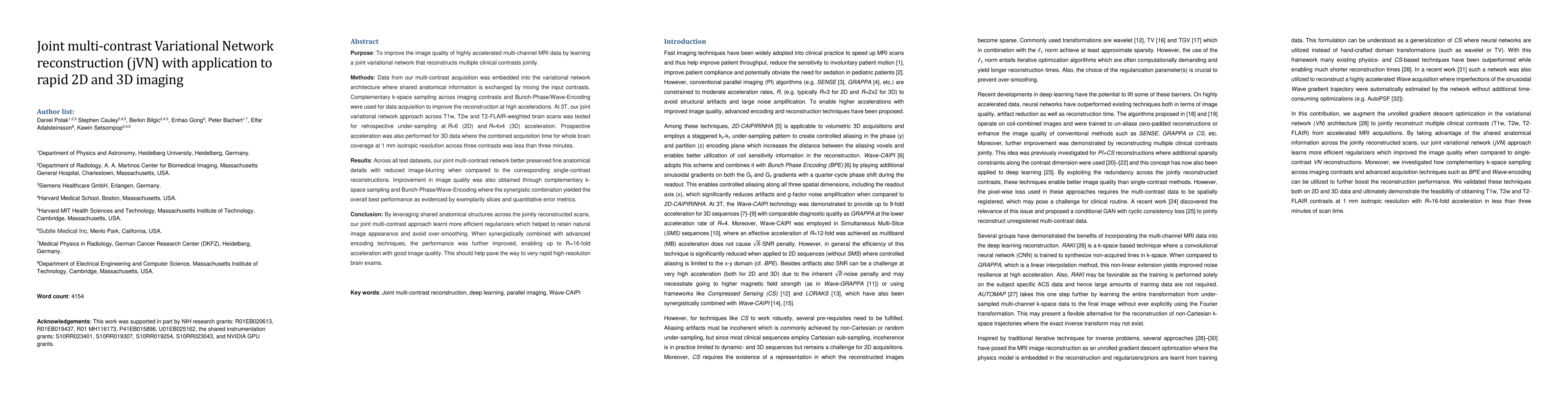
Purpose: To improve the image quality of highly accelerated multi-channel MRI data by learning a joint variational network that reconstructs multiple clinical contrasts jointly. Methods: Data from our multi-contrast acquisition was embedded into the variational network architecture where shared anatomical information is exchanged by mixing the input contrasts. Complementary k-space sampling across imaging contrasts and Bunch-Phase/Wave-Encoding were used for data acquisition to improve the reconstruction at high accelerations. At 3T, our joint variational network approach across T1w, T2w and T2-FLAIR-weighted brain scans was tested for retrospective under-sampling at R=6 (2D) and R=4x4 (3D) acceleration. Prospective acceleration was also performed for 3D data where the combined acquisition time for whole brain coverage at 1 mm isotropic resolution across three contrasts was less than three minutes. Results: Across all test datasets, our joint multi-contrast network better preserved fine anatomical details with reduced image-blurring when compared to the corresponding single-contrast reconstructions. Improvement in image quality was also obtained through complementary k-space sampling and Bunch-Phase/Wave-Encoding where the synergistic combination yielded the overall best performance as evidenced by exemplarily slices and quantitative error metrics. Conclusion: By leveraging shared anatomical structures across the jointly reconstructed scans, our joint multi-contrast approach learnt more efficient regularizers which helped to retain natural image appearance and avoid over-smoothing. When synergistically combined with advanced encoding techniques, the performance was further improved, enabling up to R=16-fold acceleration with good image quality. This should help pave the way to very rapid high-resolution brain exams.
AI Key Findings
Get AI-generated insights about this paper's methodology, results, significance, and more — seven facets brought into focus.
Impact
Paper Details
Authors
PDF Preview
Key Terms
Citation Network
Current paper (gray), citations (green), references (blue)
Display is limited for performance on very large graphs.
Discussion 0