Joint Shape Representation and Classification for Detecting PDAC
Publication
Metrics
AI Quick Summary
This paper proposes a two-stage framework for detecting pancreatic ductal adenocarcinoma (PDAC) in CT scans by segmenting the pancreas and compressing it into a shape vector for joint classification. The method achieves 90.2% specificity and 80.2% sensitivity, indicating potential for early pancreatic cancer diagnosis.
Paper Preview
Abstract
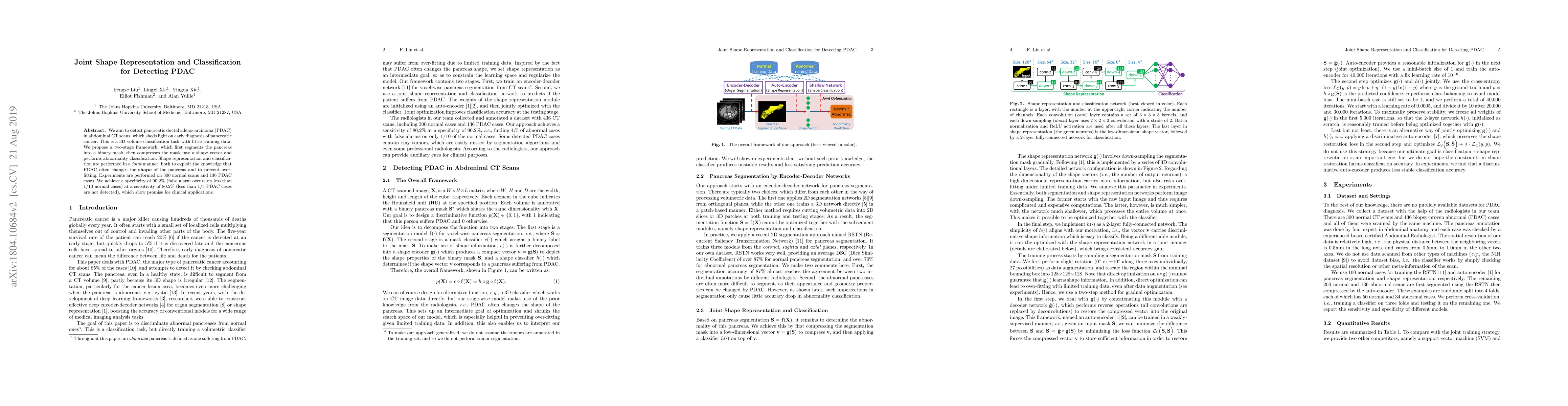
We aim to detect pancreatic ductal adenocarcinoma (PDAC) in abdominal CT scans, which sheds light on early diagnosis of pancreatic cancer. This is a 3D volume classification task with little training data. We propose a two-stage framework, which first segments the pancreas into a binary mask, then compresses the mask into a shape vector and performs abnormality classification. Shape representation and classification are performed in a joint manner, both to exploit the knowledge that PDAC often changes the shape of the pancreas and to prevent over-fitting. Experiments are performed on 300 normal scans and 136 PDAC cases. We achieve a specificity of 90.2% (false alarm occurs on less than 1/10 normal cases) at a sensitivity of 80.2% (less than 1/5 PDAC cases are not detected), which show promise for clinical applications.
AI Key Findings
Get AI-generated insights about this paper's methodology, results, significance, and more — seven facets brought into focus.
Impact
Paper Details
Authors
PDF Preview
Key Terms
Citation Network
Current paper (gray), citations (green), references (blue)
Display is limited for performance on very large graphs.
Discussion 0