Quick Answers
What methodology did the authors use?
The research employs a 3D latent diffusion model combining DDPMs, DDIMs, and VQ-GANs for weakly supervised kidney anomaly detection using contrast-enhanced abdominal CT scans. Pseudo-labels are generated from radiology reports, and post-processing techniques are applied to refine anomaly maps. More in Methodology →
What are the key results?
The method achieved lower segmentation performance compared to supervised baselines (DSC 0.08-0.12 vs. 0.68 for nnU-Net) — DDIM failed to detect lesions ≥4cm, while DDPM failed for lesions ≥7cm More in Key Results →
Why is this work significant?
This work advances 3D generative modeling for abdominal anatomy, offering annotation-efficient approaches for anomaly detection despite current performance gaps. It provides insights into diffusion-based reconstruction challenges in complex anatomical regions. More in Significance →
What are the main limitations?
Current results do not match supervised baselines — Anatomical reconstruction artifacts caused false positives More in Limitations →
Paper Preview
Abstract
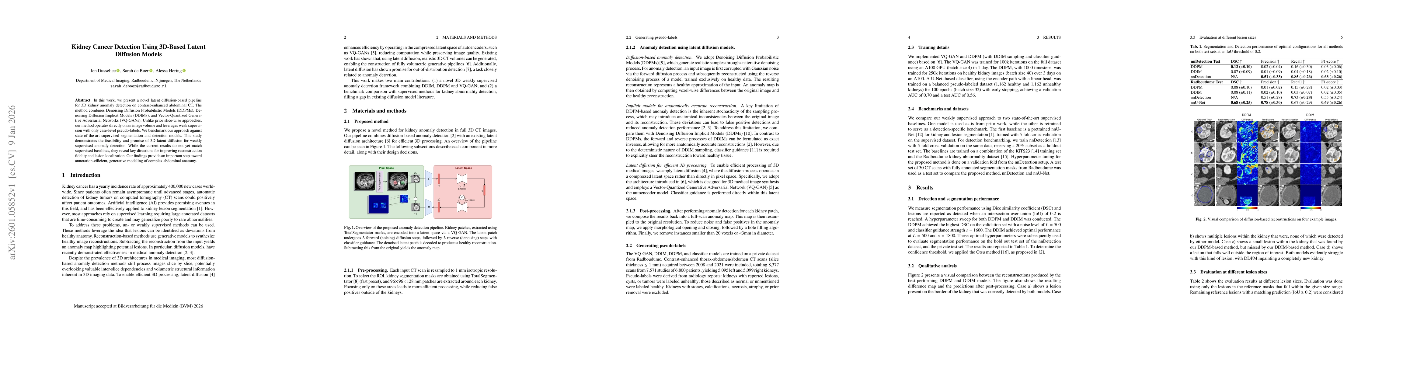
In this work, we present a novel latent diffusion-based pipeline for 3D kidney anomaly detection on contrast-enhanced abdominal CT. The method combines Denoising Diffusion Probabilistic Models (DDPMs), Denoising Diffusion Implicit Models (DDIMs), and Vector-Quantized Generative Adversarial Networks (VQ-GANs). Unlike prior slice-wise approaches, our method operates directly on an image volume and leverages weak supervision with only case-level pseudo-labels. We benchmark our approach against state-of-the-art supervised segmentation and detection models. This study demonstrates the feasibility and promise of 3D latent diffusion for weakly supervised anomaly detection. While the current results do not yet match supervised baselines, they reveal key directions for improving reconstruction fidelity and lesion localization. Our findings provide an important step toward annotation-efficient, generative modeling of complex abdominal anatomy.
AI Key Findings
Generated Jan 12, 2026
Methodology — What approach did the authors take?
The research employs a 3D latent diffusion model combining DDPMs, DDIMs, and VQ-GANs for weakly supervised kidney anomaly detection using contrast-enhanced abdominal CT scans. Pseudo-labels are generated from radiology reports, and post-processing techniques are applied to refine anomaly maps.
Key Results — What are the main findings?
- The method achieved lower segmentation performance compared to supervised baselines (DSC 0.08-0.12 vs. 0.68 for nnU-Net)
- DDIM failed to detect lesions ≥4cm, while DDPM failed for lesions ≥7cm
- Post-processing reduced false positives but risked removing smaller lesions
Significance — Why does this research matter?
This work advances 3D generative modeling for abdominal anatomy, offering annotation-efficient approaches for anomaly detection despite current performance gaps. It provides insights into diffusion-based reconstruction challenges in complex anatomical regions.
Technical Contribution — What is the technical contribution?
Development of a 3D latent diffusion framework integrating DDPM, DDIM, and VQ-GAN for weakly supervised anomaly detection in medical imaging
Novelty — What is new about this work?
Direct 3D volume processing instead of slice-wise approaches, combined with weak supervision using case-level pseudo-labels and classifier guidance within latent space
Limitations — What are the limitations of this study?
- Current results do not match supervised baselines
- Anatomical reconstruction artifacts caused false positives
- Limited performance on larger lesions
Future Work — What did the authors propose for future work?
- Investigate false positive reduction networks
- Explore alternative diffusion architectures like wavelet diffusion
- Improve guidance mechanisms for better lesion localization
Paper Details
How to Cite This Paper
@article{hering2026kidney,
  title         = {Kidney Cancer Detection Using 3D-Based Latent Diffusion Models},
  author        = {Hering, Alessa and Dusseljee, Jen and Boer, Sarah de},
  year          = {2026},
  eprint        = {2601.05852},
  archivePrefix = {arXiv},
  primaryClass  = {cs.CV},
}Hering, A., Dusseljee, J., & Boer, S. (2026). Kidney Cancer Detection Using 3D-Based Latent Diffusion Models. arXiv. https://arxiv.org/abs/2601.05852Hering, Alessa, et al. "Kidney Cancer Detection Using 3D-Based Latent Diffusion Models." arXiv, 2026, arxiv.org/abs/2601.05852.PDF Preview
Similar Papers
Found 4 papersAutoDecoding Latent 3D Diffusion Models
Evangelos Ntavelis, Aliaksandr Siarohin, Kyle Olszewski et al.
3D Cardiac Anatomy Generation Using Mesh Latent Diffusion Models
Jolanta Mozyrska, Marcel Beetz, Luke Melas-Kyriazi et al.
Unsupervised 3D out-of-distribution detection with latent diffusion models
Mark S. Graham, Walter Hugo Lopez Pinaya, Paul Wright et al.
3D Multiphase Heterogeneous Microstructure Generation Using Conditional Latent Diffusion Models
Nirmal Baishnab, Ethan Herron, Aditya Balu et al.
Comments (0)