Publication
Metrics
AI Quick Summary
This paper proposes a novel 3D U-Net framework enhanced with Expectation Maximization to segment kidneys in preclinical MRI, addressing challenges like small sample sizes and anisotropic resolution. The method achieved a Dice similarity coefficient of 0.88 on a dataset of 196 images, demonstrating its potential for various renal injuries.
Paper Preview
Abstract
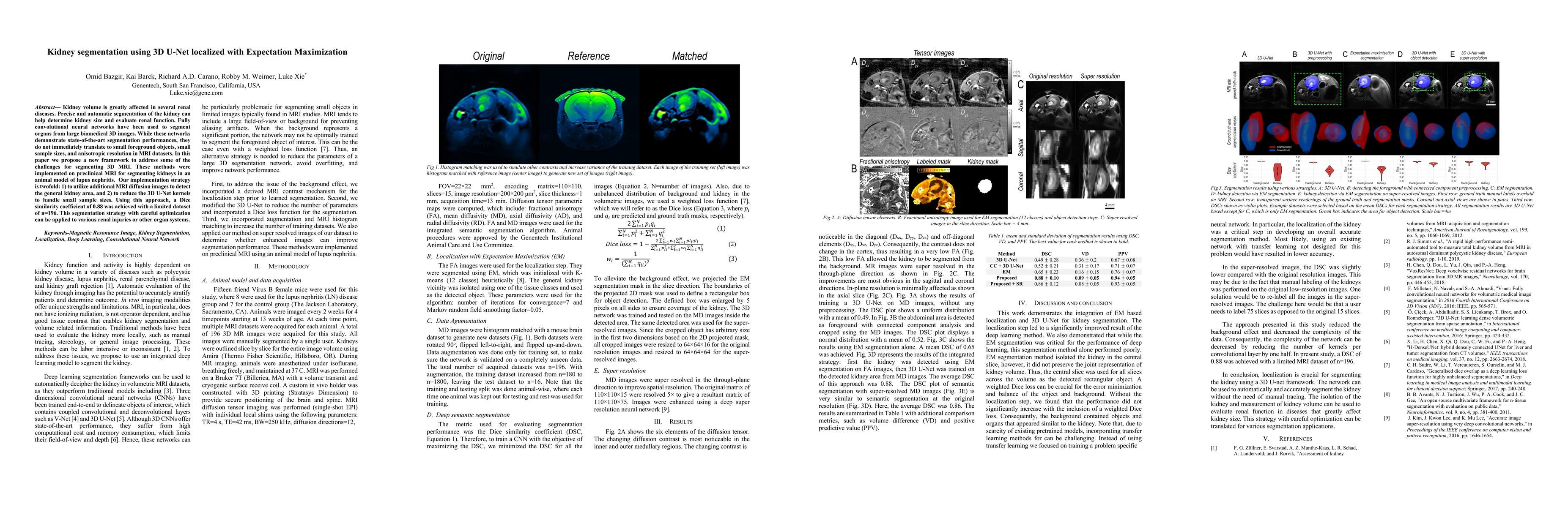
Kidney volume is greatly affected in several renal diseases. Precise and automatic segmentation of the kidney can help determine kidney size and evaluate renal function. Fully convolutional neural networks have been used to segment organs from large biomedical 3D images. While these networks demonstrate state-of-the-art segmentation performances, they do not immediately translate to small foreground objects, small sample sizes, and anisotropic resolution in MRI datasets. In this paper we propose a new framework to address some of the challenges for segmenting 3D MRI. These methods were implemented on preclinical MRI for segmenting kidneys in an animal model of lupus nephritis. Our implementation strategy is twofold: 1) to utilize additional MRI diffusion images to detect the general kidney area, and 2) to reduce the 3D U-Net kernels to handle small sample sizes. Using this approach, a Dice similarity coefficient of 0.88 was achieved with a limited dataset of n=196. This segmentation strategy with careful optimization can be applied to various renal injuries or other organ systems.
AI Key Findings
Get AI-generated insights about this paper's methodology, results, significance, and more — seven facets brought into focus.
Impact
Paper Details
Authors
PDF Preview
Key Terms
Citation Network
Current paper (gray), citations (green), references (blue)
Display is limited for performance on very large graphs.
Discussion 0