KLDD: Kalman Filter based Linear Deformable Diffusion Model in Retinal Image Segmentation
Publication
Metrics
AI Quick Summary
This paper proposes a Kalman Filter based Linear Deformable Diffusion (KLDD) model for retinal vessel segmentation, aiming to better capture small blood vessels. The KLDD model uses deformable convolution and a Kalman filter for feature extraction and optimization, followed by Cross-Attention Aggregation and Channel-wise Soft Attention modules to refine segmentation, showing superior performance on multiple retinal image datasets.
Paper Preview
Abstract
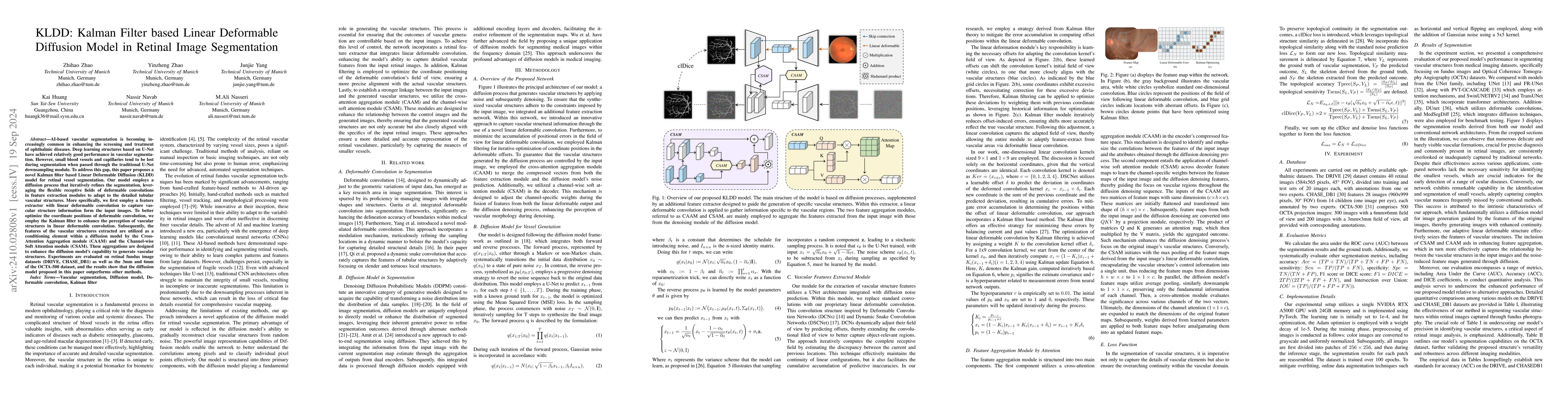
AI-based vascular segmentation is becoming increasingly common in enhancing the screening and treatment of ophthalmic diseases. Deep learning structures based on U-Net have achieved relatively good performance in vascular segmentation. However, small blood vessels and capillaries tend to be lost during segmentation when passed through the traditional U-Net downsampling module. To address this gap, this paper proposes a novel Kalman filter based Linear Deformable Diffusion (KLDD) model for retinal vessel segmentation. Our model employs a diffusion process that iteratively refines the segmentation, leveraging the flexible receptive fields of deformable convolutions in feature extraction modules to adapt to the detailed tubular vascular structures. More specifically, we first employ a feature extractor with linear deformable convolution to capture vascular structure information form the input images. To better optimize the coordinate positions of deformable convolution, we employ the Kalman filter to enhance the perception of vascular structures in linear deformable convolution. Subsequently, the features of the vascular structures extracted are utilized as a conditioning element within a diffusion model by the Cross-Attention Aggregation module (CAAM) and the Channel-wise Soft Attention module (CSAM). These aggregations are designed to enhance the diffusion model's capability to generate vascular structures. Experiments are evaluated on retinal fundus image datasets (DRIVE, CHASE_DB1) as well as the 3mm and 6mm of the OCTA-500 dataset, and the results show that the diffusion model proposed in this paper outperforms other methods.
AI Key Findings
Get AI-generated insights about this paper's methodology, results, significance, and more — seven facets brought into focus.
Impact
Paper Details
Authors
PDF Preview
Citation Network
Current paper (gray), citations (green), references (blue)
Display is limited for performance on very large graphs.
Discussion 0