Knowledge-Augmented Contrastive Learning for Abnormality Classification and Localization in Chest X-rays with Radiomics using a Feedback Loop
Publication
Metrics
AI Quick Summary
The approach leverages a feedback loop of image and radiomic features to enhance robustness and interpretability of the model.
Paper Preview
Abstract
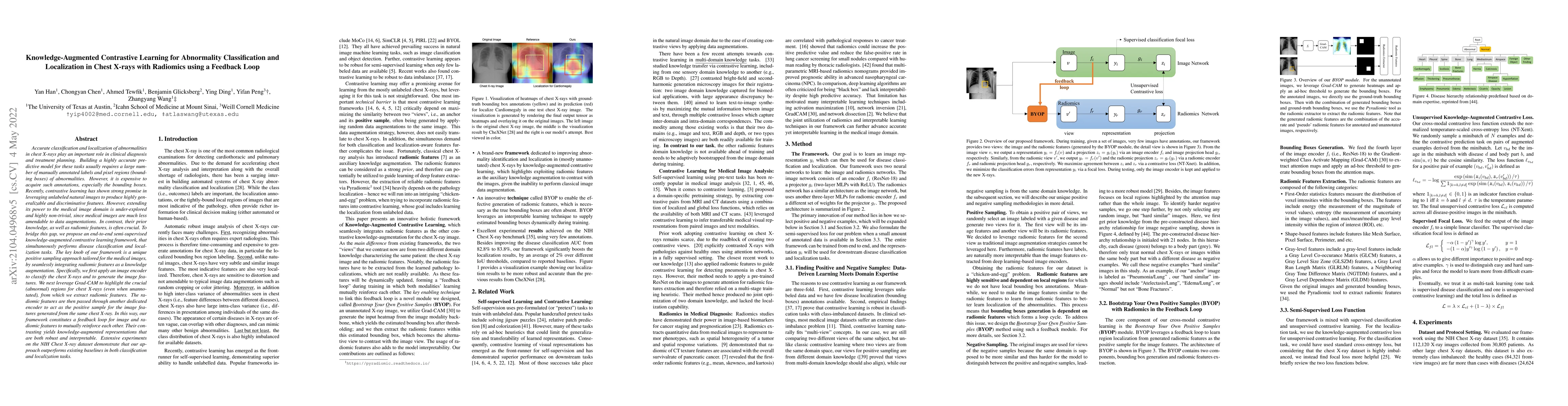
Building a highly accurate predictive model for classification and localization of abnormalities in chest X-rays usually requires a large number of manually annotated labels and pixel regions (bounding boxes) of abnormalities. However, it is expensive to acquire such annotations, especially the bounding boxes. Recently, contrastive learning has shown strong promise in leveraging unlabeled natural images to produce highly generalizable and discriminative features. However, extending its power to the medical image domain is under-explored and highly non-trivial, since medical images are much less amendable to data augmentations. In contrast, their prior knowledge, as well as radiomic features, is often crucial. To bridge this gap, we propose an end-to-end semi-supervised knowledge-augmented contrastive learning framework, that simultaneously performs disease classification and localization tasks. The key knob of our framework is a unique positive sampling approach tailored for the medical images, by seamlessly integrating radiomic features as a knowledge augmentation. Specifically, we first apply an image encoder to classify the chest X-rays and to generate the image features. We next leverage Grad-CAM to highlight the crucial (abnormal) regions for chest X-rays (even when unannotated), from which we extract radiomic features. The radiomic features are then passed through another dedicated encoder to act as the positive sample for the image features generated from the same chest X-ray. In this way, our framework constitutes a feedback loop for image and radiomic modality features to mutually reinforce each other. Their contrasting yields knowledge-augmented representations that are both robust and interpretable. Extensive experiments on the NIH Chest X-ray dataset demonstrate that our approach outperforms existing baselines in both classification and localization tasks.
AI Key Findings
Get AI-generated insights about this paper's methodology, results, significance, and more — seven facets brought into focus.
Impact
Paper Details
Authors
PDF Preview
Key Terms
Citation Network
Current paper (gray), citations (green), references (blue)
Display is limited for performance on very large graphs.
Discussion 0