This paper presents a label-free method using optical diffraction tomography to characterize white blood cells (WBCs) by constructing 3D refractive index maps, enabling the separation of WBC populations based on quantitative morphological and biochemical data. The technique also tracks the 3D trajectory of ingested micrometer-sized beads, showcasing its potential for WBC analysis.
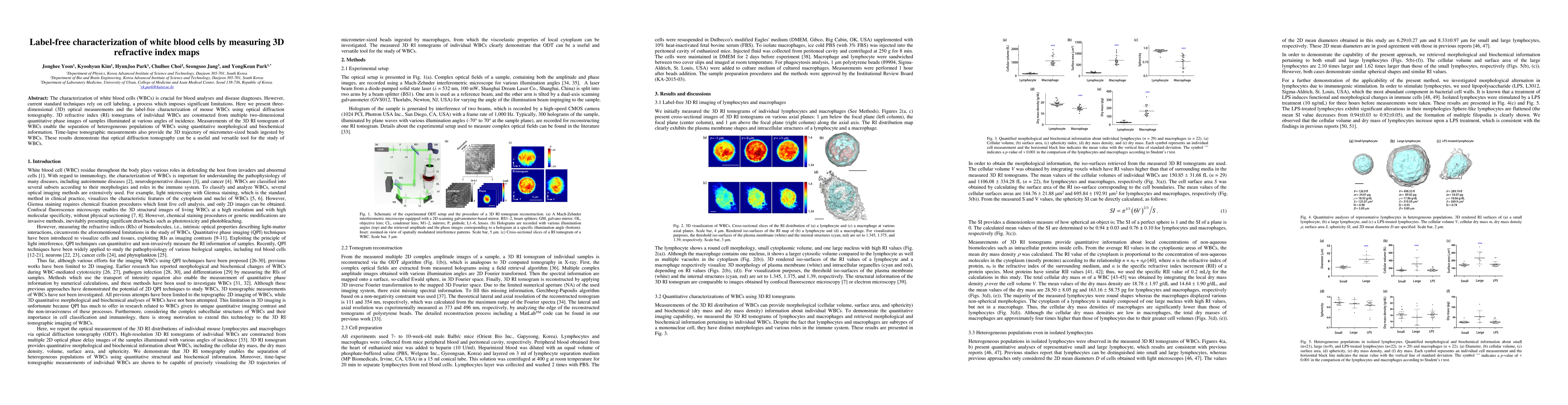
The characterization of white blood cells (WBCs) is crucial for blood analyses and disease diagnoses. However, current standard techniques rely on cell labeling, a process which imposes significant limitations. Here we present three-dimensional (3D) optical measurements and the label-free characterization of mouse WBCs using optical diffraction tomography. 3D refractive index (RI) tomograms of individual WBCs are constructed from multiple two-dimensional quantitative phase images of samples illuminated at various angles of incidence. Measurements of the 3D RI tomogram of WBCs enable the separation of heterogeneous populations of WBCs using quantitative morphological and biochemical information. Time-lapse tomographic measurements also provide the 3D trajectory of micrometer-sized beads ingested by WBCs. These results demonstrate that optical diffraction tomography can be a useful and versatile tool for the study of WBCs.
Get AI-generated insights about this paper's methodology, results, and significance.
Current paper (gray), citations (green), references (blue)
Rohit Bhargava, Jaena Park, Krishna Agarwal et al.
Title | Authors | Year | Actions |
---|---|---|---|
Saccharomyces cerevisiae recovery from various mild abiotic stresses: Viability, fitness, and high resolution three-dimensional morphology imaging.
|
Piotr J. Pietras, Monika Chaszczewska-Markowska, Daniel Ghete, Agata Tyczewska, K. Bąkowska-Żywicka
|
2025
|
View Paper |
Differential sensitivity of hemocyte subpopulations (Mytilus edulis) to aged polyethylene terephthalate micro- and nanoplastic particles.
|
Jenevieve Hara, M. Vercauteren, Sébastjen Schoenaers, Colin Janssen, Ronny Blust, J. Asselman, R. Town
|
2024
|
View Paper |
Azadirachta indica fruit juice clears malaria parasites and replenishes blood levels in Plasmodium berghei-infected mice
|
Igwenyi Ikechuku Okorie, Onodugo Chinemelum Adaora, Aja Patrick Maduabuchi, E. Oge, Awoke Joshua Nonso, Ibhadode Osarieme Sandra, Uraku Anayo Joseph, Edwin Nzubechukwu, Obasi Ajuka Nwogo, David Ebuka Elijah, Igwenyi Ijeoma Precious, Ogbu Patience Nkemjika, Egwu Chinedu, Atoki Ayomide Victor
|
2024
|
View Paper |
A highly sensitive modified triple split ring metamaterial-based sensor for blood sample detection based on dielectric property alteration
|
Abdullah Al Mahfazur Rahman, Mohammad Tariqul Islam, Phumin Kirawanich, Badariah Bais, Haitham Alsaif, Abdulwadoud A. Maash, Ahasanul Hoque, Md. Moniruzzaman, M. Islam, Mohamed S. Soliman
|
2024
|
View Paper |
Quantitative Phase Imaging in Tissue with Gradient Retardance Optical Microscopy
|
Jinming Zhang, M. Sarollahi, Andreas E. Vasdekis
|
2024
|
View Paper |
Unified, Verifiable Neural Simulators for Electromagnetic Wave Inverse Problems
|
Charles Dove, Jatearoon Boondicharern, Laura Waller
|
2024
|
View Paper |
Imaging the intracellular refractive index distribution (IRID) for dynamic label-free living colon cancer cells via circularly depolarization decay model (CDDM)
|
Huijun Wang, Lu Zhang, Jie Huang, Zewen Yang, Chen Fan, Li Yuan, Hongwuyi Zhao, Zhenxi Zhang, Xiaolong Liu
|
2024
|
View Paper |
High-precision and low-noise dielectric tensor tomography using a micro-electromechanical system mirror.
|
Juheon Lee, Byung Gyu Chae, Hyun-Eui Kim, MinSung Yoon, Hervé Hugonnet, YongKeun Park
|
2024
|
View Paper |
Deep learning-assisted smartphone-based quantitative microscopy for label-free peripheral blood smear analysis
|
Bingxin Huang, Lei Kang, V. C. Tsang, Claudia T. K. Lo, Terence T. W. Wong
|
2024
|
View Paper |
Stimulated Raman scattering microscopy in the analysis of cancer cells – A review and own research
|
A. Adamczyk, Sylwia Orzechowska, A. Nowakowska, Krzysztof Brzozowski, K. Majzner, M. Baranska
|
2023
|
View Paper |
Holo-tomographic flow cytometry: a new paradigm in diagnostics by high-throughput and stain-free single-cell imaging
|
L. Miccio, D. Pirone, J. Běhal, G. Giugliano, Michela Schiavo, V. Bianco, P. Memmolo, P. Ferraro
|
2023
|
View Paper |
Non–steady state thermometry with optical diffraction tomography
|
Adarsh B. Vasista, B. Ciraulo, Falko Schmidt, J. O. Arroyo, R. Quidant
|
2023
|
View Paper |
Deep Learning Assisted Smartphone-based Quantitative Microscopy for Label-free Hematological Analysis
|
Bingxin Huang, Lei Kang, V. C. Tsang, Claudia T. K. Lo, T. T. Wong
|
2023
|
View Paper |
Stain-free identification of cell nuclei using tomographic phase microscopy in flow cytometry
|
D. Pirone, Joowon Lim, F. Merola, L. Miccio, M. Mugnano, V. Bianco, F. Cimmino, Feliciano Visconte, Annalaura Montella, M. Capasso, A. Iolascon, P. Memmolo, D. Psaltis, P. Ferraro
|
2022
|
View Paper |
Developing a Reliable Holographic Flow Cyto-Tomography Apparatus by Optimizing the Experimental Layout and Computational Processing
|
J. Běhal, F. Borrelli, M. Mugnano, V. Bianco, A. Capozzoli, C. Curcio, A. Liseno, L. Miccio, P. Memmolo, P. Ferraro
|
2022
|
View Paper |
Diagnosis of Unstained Biological Blood Cells Using Phase Hologram
|
D. Ibrahim
|
2022
|
View Paper |
Functional blood cell analysis by label-free biosensors and single-cell technologies.
|
Zoltán Szittner, B. Péter, S. Kurunczi, I. Székács, R. Horváth
|
2022
|
View Paper |
Deep Learning of Morphologic Correlations To Accurately Classify CD4+ and CD8+ T Cells by Diffraction Imaging Flow Cytometry.
|
Lin Zhao, Liwen Tang, Marion S Greene, Y. Sa, Wenjin Wang, Jiahong Jin, Heng Hong, Jun Q. Lu, Xin-Hua Hu
|
2022
|
View Paper |
Compact common-path polarisation holographic microscope for measuring spatially-resolved Jones matrix parameters of dynamic samples in microfluidics
|
Guiying Chen, Yingying Zhang, Yang Yang, Hong-Yi Huang, Qing-yang Yue, Cheng-Shan Guo
|
2022
|
View Paper |
Stain-free nucleus identification in holographic learning flow cyto-tomography
|
D. Pirone, Joowon Lim, F. Merola, L. Miccio, M. Mugnano, V. Bianco, F. Cimmino, Feliciano Visconte, Annalaura Montella, M. Capasso, A. Iolascon, P. Memmolo, D. Psaltis, P. Ferraro
|
2021
|
View Paper |
Label-free imaging and evaluation of characteristic properties of asthma-derived eosinophils using optical diffraction tomography.
|
Sang‐Yeob Kim, Ji-Hyang Lee, Yeonhee Shin, Tae-Keun Kim, Ji won Lee, Minju Pyo, Amanda R. Lee, C. Pack, Y. Cho
|
2021
|
View Paper |
Label-free tomographic imaging of nanodiamonds in living cells
|
Dini Kurnia Ikliptikawati, M. Hazawa, Frederick T.-K. So, Daiki Terada, Akiko Kobayashi, T. Segawa, M. Shirakawa, R. Wong
|
2021
|
View Paper |
Label-free automated neutropenia detection and grading using deep-ultraviolet microscopy.
|
A. Ojaghi, Paloma Casteleiro Costa, C. Caruso, W. Lam, Francisco E. Robles
|
2021
|
View Paper |
Autofluorescence of blood and its application in biomedical and clinical research
|
A. Shrirao, R. Schloss, Zachary R. Fritz, Mayur V. Shrirao, Robert Rosen, M. Yarmush
|
2021
|
View Paper |
Optimization analysis of partially coherent illumination for refractive index tomographic microscopy
|
Jiaji Li, Ning Zhou, Zhidong Bai, Shun Zhou, Qian Chen, Chao Zuo
|
2021
|
View Paper |
Reagent- and actuator-free analysis of individual erythrocytes using three-dimensional quantitative phase imaging and capillary microfluidics
|
DongHun Ryu, Hyeono Nam, J. Jeon, Yongkeun Park
|
2021
|
View Paper |
Detection of intracellular monosodium urate crystals in gout synovial fluid using optical diffraction tomography
|
Sangwoo Park, L. E. Lee, Hanna Kim, J. Kim, Seung Jun Lee, S. Yoon, Seungwoo Shin, H. Kang, Yongkeun Park, J. Song, Seongsoo Lee
|
2021
|
View Paper |
Limited-angle tomographic phase microscopy utilizing confocal scanning fluorescence microscopy.
|
Rongli Guo, I. Barnea, N. Shaked
|
2021
|
View Paper |
Holographic tomography: techniques and biomedical applications [Invited].
|
Vinoth Balasubramani, A. Kuś, H. Tu, Chau-Jern Cheng, M. Baczewska, W. Krauze, M. Kujawińska
|
2021
|
View Paper |
Quantitative phase imaging trends in biomedical applications
|
T. Cacace, V. Bianco, P. Ferraro
|
2020
|
View Paper |
Label-free quantitative 3D intensity diffraction tomographic imaging in high numerical aperture microscopy
|
Jiaji Li, Alex Matlock, Yunzhe Li, Qian Chen, L. Tian, Chao Zuo
|
2020
|
View Paper |
3D resolution-enhanced intensity diffraction tomographic microscopy
|
Jiaji Li, Alex Matlock, Yunzhe Li, Qian Chen, L. Tian, Chao Zuo
|
2020
|
View Paper |
Multi-pulse laser-induced bubble formation and nanoparticle aggregation using MoS2 nanoparticles
|
B. Ko, Weigang Lu, A. Sokolov, H. W. Lee, M. Scully, Zhenrong Zhang
|
2020
|
View Paper |
Fast compressive lens-free tomography for 3D biological cell culture imaging.
|
Zhenxiang Luo, A. Yurt, R. Stahl, M. Carlon, A. Ramalho, F. Vermeulen, A. Lambrechts, D. Braeken, L. Lagae
|
2020
|
View Paper |
Label-free hematology analysis using deep-ultraviolet microscopy
|
A. Ojaghi, G. Carrazana, C. Caruso, Asad Abbas, D. Myers, W. Lam, Francisco E. Robles
|
2020
|
View Paper |
Micromirror-Embedded Coverslip Assembly for Bidirectional Microscopic Imaging
|
Dongwoo Lee, Jihye Kim, E. Song, Ji-young Jeong, E. Jeon, Pilhan Kim, Wonhee Lee
|
2020
|
View Paper |
3D morphological and biophysical changes in a single tachyzoite and its infected cells using three‐dimensional quantitative phase imaging
|
Egy Rahman Firdaus, Ji-Hoon Park, Seong-kyun Lee, Yongkeun Park, Guang-Ho Cha, E. Han
|
2020
|
View Paper |
Real-time cholesterol sorting in Plasmodium falciparum-erythrocytes as revealed by 3D label-free imaging
|
E. Hayakawa, K. Yamaguchi, M. Mori, G. Nardone
|
2020
|
View Paper |
Computational microscopy for quantitative phase imaging and refractive index tomography using annular illumination
|
Jiaji Li, Alex Matlock, Yunzhe Li, Qian Chen, L. Tian, Chao Zuo
|
2020
|
View Paper |
Label-free three-dimensional observations and quantitative characterisation of on-chip vasculogenesis using optical diffraction tomography
|
Chungha Lee, Seunggyu Kim, Hervé Hugonnet, Moosung Lee, W. Park, J. Jeon, Yongkeun Park
|
2020
|
View Paper |
Measurements of 3-D morphology of individual red blood cells upon localized photothermal effects using gold nanorods
|
Chan Song, Gu-Haeng Lee, Juntaek Oh, Moosung Lee, Seungwoo Shin, Sangyun Lee, Y. Nam, Yongkeun Park
|
2019
|
View Paper |
Impact of sample preparation on holographic imaging of leukocytes
|
C. Klenk, D. Heim, Matthias Ugele, O. Hayden
|
2019
|
View Paper |
High-speed in vitro intensity diffraction tomography
|
Jiaji Li, Alex Matlock, Yunzhe Li, Qian Chen, Chao Zuo, L. Tian
|
2019
|
View Paper |
Versatile transmission/reflection tomographic diffractive microscopy approach.
|
L. Foucault, N. Verrier, M. Debailleul, Jean-Baptiste Courbot, B. Colicchio, B. Simon, L. Vonna, O. Haeberlé
|
2019
|
View Paper |
High-fidelity optical diffraction tomography of multiple scattering samples
|
Joowon Lim, Ahmed B. Ayoub, Elizabeth E. Antoine, D. Psaltis
|
2019
|
View Paper |
An exploratory study towards objective quality evaluation of digital hologram coding tools
|
Roberto Corda, Antonin Gilles, Kwan‐Jung Oh, António M. G. Pinheiro, P. Schelkens, C. Perra
|
2019
|
View Paper |
Label-free characterization of white blood cells using fluorescence lifetime imaging and flow-cytometry: molecular heterogeneity and erythrophagocytosis [Invited].
|
B. P. Yakimov, M. Gogoleva, A. Semenov, S. A. Rodionov, M. Novoselova, A. Gayer, A. V. Kovalev, A. I. Bernakevich, V. Fadeev, Artashes G. Armaganov, V. Drachev, D. Gorin, M. Darvin, V. Shcheslavskiy, Gleb S. Budylin, A. Priezzhev, E. Shirshin
|
2019
|
View Paper |
Quantitative phase imaging in common-path cross-referenced holographic microscopy using double-exposure method
|
J. Běhal
|
2019
|
View Paper |
Advanced label-free cellular identification in flow by collaborative coherent imaging techniques
|
D. Dannhauser, Domenico Rossi, Maria Isabella Maremonti, P. Memmolo, P. Ferraro, P. Netti, F. Causa
|
2019
|
View Paper |
Ocular anterior chamber blood cell population differentiation using spectroscopic optical coherence tomography.
|
Ruobing Qian, Weifeng Huang, R. McNabb, Kevin C Zhou, Q. Liu, A. Kuo, J. Izatt
|
2019
|
View Paper |
Statistical analysis of computer generated holography for data compression applications
|
Roberto Corda, D. Giusto, C. Perra, A. Liotta, Wei Song
|
2019
|
View Paper |
A method for assessing the fidelity of optical diffraction tomography reconstruction methods using structured illumination
|
Ahmed B. Ayoub, Thanh-an Michel Pham, Joowon Lim, M. Unser, D. Psaltis
|
2019
|
View Paper |
Recent Advances in the Processing and Rendering Algorithms for Computer-Generated Holography
|
Roberto Corda, D. Giusto, A. Liotta, Wei Song, C. Perra
|
2019
|
View Paper |
Label‐free bioanalysis of Leishmania infantum using refractive index tomography with partially coherent illumination
|
J. Soto, Alicia Mas, J. Rodrigo, T. Alieva, G. Domínguez-Bernal
|
2019
|
View Paper |
High-speed in vitro intensity diffraction tomography
|
Jiaji Li, Alex Matlock, Yunzhe Li, Qian Chen, Chao Zuo, L. Tian
|
2019
|
View Paper |
Label-free classification of cells based on supervised machine learning of subcellular structures
|
Yusuke Ozaki, Hidenao Yamada, H. Kikuchi, Amane Hirotsu, T. Murakami, Tomohiro Matsumoto, Toshiki Kawabata, Y. Hiramatsu, K. Kamiya, T. Yamauchi, Kentaro Goto, Y. Ueda, S. Okazaki, M. Kitagawa, H. Takeuchi, H. Konno
|
2019
|
View Paper |
Label-free classification of neurons and glia in neural stem cell cultures using a hyperspectral imaging microscopy combined with machine learning
|
H. Ogi, Sanzo Moriwaki, M. Kokubo, Yuichiro Hikida, K. Itoh
|
2019
|
View Paper |
Cycle-consistent deep learning approach to coherent noise reduction in optical diffraction tomography.
|
Gunho Choi, DongHun Ryu, YoungJu Jo, Youngseo Kim, W. Park, Hyun-Seok Min, Yongkeun Park
|
2018
|
View Paper |
Enhancement of optical resolution in three-dimensional refractive-index tomograms of biological samples by employing micromirror-embedded coverslips.
|
Seungwoo Shin, Jihye Kim, Je-Ryung Lee, E. Jeon, T. Je, Wonhee Lee, Yongkeun Park
|
2018
|
View Paper |
Recent Advancements and Perspective About Digital Holography: A Super-Tool in Biomedical and Bioengineering Fields
|
F. Merola, B. Mandracchia, L. Miccio, P. Memmolo, V. Bianco, M. Mugnano, P. Maffettone, M. Villone, E. Maio, V. Ferraro, Zhe Wang, V. Pagliarulo, S. Grilli, P. Ferraro
|
2018
|
View Paper |
Axonal Transport, Phase-Separated Compartments, and Neuron Mechanics - A New Approach to Investigate Neurodegenerative Diseases
|
M. Nötzel, Gonzalo Rosso, Stephanie Möllmert, Anne Seifert, Raimund Schlüßler, Kyoohyun Kim, A. Hermann, J. Guck
|
2018
|
View Paper |
Biophysical investigation of living monocytes in flow by collaborative coherent imaging techniques.
|
D. Dannhauser, Domenico Rossi, P. Memmolo, A. Finizio, P. Ferraro, P. Netti, F. Causa
|
2018
|
View Paper |
Combining Three-Dimensional Quantitative Phase Imaging and Fluorescence Microscopy for the Study of Cell Pathophysiology
|
Young Seo Kim, Sangyun Lee, Jaehwang Jung, Seungwoo Shin, HeeSung Choi, Guang-Ho Cha, W. Park, Sumin Lee, Yongkeun Park
|
2018
|
View Paper |
Triplex micron-resolution acoustic, photoacoustic, and optical transmission microscopy via photoacoustic radiometry.
|
M. Moore, E. Strohm, Michael C. Kolios
|
2018
|
View Paper |
Clinical lymphocytes construction for light scattering inversion study: a three-dimensional morphology constructed method from defective confocal images
|
Lu Zhang, Yunhao Xie, Yingzhe Tu, Lele Luo, Kaixing Li, Li Yuan, Wei Chen, Hong Zhao, Zhenxi Zhang
|
2018
|
View Paper |
Three-dimensional tomographic microscopy technique with multi-frequency combination with partially coherent illuminations
|
Jiaji Li, Jiasong Sun, Jialin Zhang, Qian Chen, Chao Zuo
|
2018
|
View Paper |
Label-free quantitative evaluation of breast tissue using Spatial Light Interference Microscopy (SLIM)
|
Hassaan Majeed, Tan H. Nguyen, M. Kandel, A. Kajdacsy-Balla, G. Popescu
|
2018
|
View Paper |
Three-dimensional optical diffraction tomographic microscopy with optimal frequency combination with partially coherent illuminations
|
Jiaji Li, Qianzhen Sun, Jialin Zhang, Junyi Ding, Chao Zuo
|
2018
|
View Paper |
Phase contrast tomography at lab on chip scale by digital holography.
|
F. Merola, P. Memmolo, L. Miccio, M. Mugnano, P. Ferraro
|
2018
|
View Paper |
Digital holography and its multidimensional imaging applications: a review
|
T. Tahara, X. Quan, R. Otani, Y. Takaki, O. Matoba
|
2018
|
View Paper |
Advanced multimodal methods in biomedicine : Raman spectroscopy and digital holographic microscopy
|
Naomi McReynolds
|
2017
|
View Paper |
Correlative three-dimensional fluorescence and refractive index tomography: bridging the gap between molecular specificity and quantitative bioimaging.
|
Kyoohyun Kim, W. Park, Sangchan Na, Sangbum Kim, Taehong Kim, Won Do Heo, Yongkeun Park
|
2017
|
View Paper |
Label-free non-invasive quantitative measurement of lipid contents in individual microalgal cells using refractive index tomography
|
Jaehwang Jung, Seong-Joo Hong, Han-Byeol Kim, Geon Kim, Moosung Lee, Seungwoo Shin, Sangyun Lee, Dong-Jin Kim, Choul-gyun Lee, Yongkeun Park
|
2017
|
View Paper |
Label-free quantitative screening of breast tissue using Spatial Light Interference Microscopy (SLIM)
|
Hassaan Majeed, Tan H. Nguyen, M. Kandel, A. Kajdacsy-Balla, G. Popescu
|
2017
|
View Paper |
Image quality improvement in optical diffraction tomography by multiple numerical propagations and separated reconstructions
|
Xichao Ma, Wen Xiao, Feng Pan
|
2017
|
View Paper |
Measurements of three-dimensional refractive index tomography and membrane deformability of live erythrocytes from Pelophylax nigromaculatus
|
Geon Kim, Moosung Lee, SeongYeon Youn, EuiTae Lee, Daeheon Kwon, Jonghun Shin, Sangyun Lee, Youn Sil Lee, Yongkeun Park
|
2017
|
View Paper |
Correlative three-dimensional fluorescence and refractive index tomography: bridging the gap between molecular specificity and quantitative bioimaging
|
Kyoohyun Kim, W. Park, Sangchan Na, Sangbum Kim, Taehong Kim, W. Heo, Yongkeun Park
|
2017
|
View Paper |
Quantitative analysis and comparison of 3D morphology between viable and apoptotic MCF-7 breast cancer cells and characterization of nuclear fragmentation
|
Yuhua Wen, Zhan Chen, Jianfen Lu, Elizabeth Ables, J. Scemama, Li V. Yang, Jun Q. Lu, Xin-Hua Hu
|
2017
|
View Paper |
Structured illumination microscopy for dual-modality 3D sub-diffraction resolution fluorescence and refractive-index reconstruction.
|
Shwetadwip Chowdhury, W. Eldridge, A. Wax, J. Izatt
|
2017
|
View Paper |
Holographic microscope slide in a spatio-temporal imaging modality for reliable 3D cell counting.
|
B. Mandracchia, V. Bianco, Zhe Wang, M. Mugnano, A. Bramanti, M. Paturzo, P. Ferraro
|
2017
|
View Paper |
Multimodal approach combining optical diffraction tomography and three-dimensional structured illumination microscopy using a digital micromirror device
|
Seungwoo Shin, Doyeon Kim, Yongkeun Park
|
2017
|
View Paper |
Identification of non-activated lymphocytes using three-dimensional refractive index tomography and machine learning
|
Jonghee Yoon, YoungJu Jo, Min-hyeok Kim, Kyoohyun Kim, Sangyun Lee, Suk-Jo Kang, Yongkeun Park
|
2017
|
View Paper |
Label-free quantitative 3D tomographic imaging for partially coherent light microscopy.
|
J. Soto, J. Rodrigo, T. Alieva
|
2017
|
View Paper |
Optical 3D imaging of subcellular dynamics in biological cultures and tissues : applications to ophthalmology and neuroscience
|
O. Thouvenin
|
2017
|
View Paper |
Measurements of morphological and biophysical alterations in individual neuron cells associated with early neurotoxic effects in Parkinson's disease
|
Su-A Yang, Jonghee Yoon, Kyoohyun Kim, Yongkeun Park
|
2017
|
View Paper |
Label‐free analysis of mononuclear human blood cells in microfluidic flow by coherent imaging tools
|
D. Dannhauser, Domenico Rossi, P. Memmolo, F. Causa, A. Finizio, P. Ferraro, P. Netti
|
2017
|
View Paper |
Tomographic phase microscopy: principles and applications in bioimaging [Invited].
|
Di Jin, R. Zhou, Z. Yaqoob, P. So
|
2017
|
View Paper |
Quantifying collagen fiber orientation in breast cancer using quantitative phase imaging
|
Hassaan Majeed, Chukwuemeka Okoro, A. Kajdacsy-Balla, K. Toussaint, G. Popescu
|
2017
|
View Paper |
Endowing a plain fluidic chip with micro-optics: a holographic microscope slide
|
V. Bianco, B. Mandracchia, V. Marchesano, V. Pagliarulo, F. Olivieri, S. Coppola, M. Paturzo, P. Ferraro
|
2017
|
View Paper |
Multimodal discrimination of immune cells using a combination of Raman spectroscopy and digital holographic microscopy
|
Naomi McReynolds, Fiona G. M. Cooke, Mingzhou Chen, S. Powis, K. Dholakia
|
2017
|
View Paper |
An Introduction to Tomographic Diffractive Microscopy: Toward High-Speed Quantitative Imaging Beyond the Abbe Limit
|
J. Bailleul, B. Simon, M. Debailleul, O. Haeberlé
|
2017
|
View Paper |
Label-free identification of non-activated lymphocytes using three-dimensional refractive index tomography and machine learning
|
Jonghee Yoon, YoungJu Jo, Min-hyeok Kim, Kyoohyun Kim, Sangyun Lee, Suk-Jo Kang, Yongkeun Park
|
2017
|
View Paper |
Holotomography: refractive index as an intrinsic imaging contrast for 3-D label-free live cell imaging
|
Doyeon Kim, Sangyun Lee, Moosung Lee, Juntaek Oh, Su-A Yang, Yongkeun Park
|
2017
|
View Paper |
Quantitative phase imaging for medical diagnosis
|
Hassaan Majeed, Shamira Sridharan, Mustafa Mir, Lihong Ma, Eunjung Min, W. Jung, G. Popescu
|
2017
|
View Paper |
Label-free high-resolution 3-D imaging of gold nanoparticles inside live cells using optical diffraction tomography
|
Doyeon Kim, Nuri Oh, Kyoohyun Kim, Sangyun Lee, C. Pack, Ji-Ho Park, Yongkeun Park
|
2016
|
View Paper |
Time-multiplexed structured illumination using a DMD for optical diffraction tomography.
|
KyeoReh Lee, Kyoohyun Kim, Geon Kim, Seungwoo Shin, Yongkeun Park
|
2016
|
View Paper |
Unwrapping free Hilbert transform-based phase retrieval method in quantitative interferometric microscopy
|
Shouyu Wang, Keding Yan, Nan Sun, Xiaolin Tian, Wei Yu, Yuanjie Li, Aihui Sun, Liang Xue
|
2016
|
View Paper |
Refractive index tomograms and dynamic membrane fluctuations of red blood cells from patients with diabetes mellitus
|
Sangyun Lee, Hyunjoo Park, Kyoohyun Kim, YongHak Sohn, S. Jang, Yongkeun Park
|
2016
|
View Paper |
Three-dimensional label-free imaging and quantification of lipid droplets in live hepatocytes
|
Kyoohyun Kim, Seoeun Lee, Jonghee Yoon, JiHan Heo, Chulhee Choi, Yongkeun Park
|
2016
|
View Paper |
Tomographic active optical trapping of arbitrarily shaped objects by exploiting 3D refractive index maps
|
Kyoohyun Kim, Yongkeun Park
|
2016
|
View Paper |
Optical Measurements of Stored Human Red Blood Cells with and Without CPDA-1
|
Sangyun Lee, Hyunjoo Park, Yongkeun Park
|
2016
|
View Paper |
3-D Quantitative Measurements of Individual Human Red Blood Cells from Diabetic Patients Employing 3-D Quantitative Phase Imaging
|
Sangyun Lee, Yongkeun Park
|
2016
|
View Paper |
Quantitative Phase Imaging of Fluid Mixing in Microfluid Chips
|
Gwangsik Park, Gwangsu Kim, Kyoohyun Kim, Yongkeun Park
|
2016
|
View Paper |
Rapid 3D Refractive‐Index Imaging of Live Cells in Suspension without Labeling Using Dielectrophoretic Cell Rotation
|
Mor Habaza, M. Kirschbaum, Christian R Guernth-Marschner, Gili Dardikman, I. Barnea, R. Korenstein, C. Duschl, N. Shaked
|
2016
|
View Paper |
Tomographic flow cytometry by digital holography
|
F. Merola, P. Memmolo, L. Miccio, Roberto Savoia, M. Mugnano, A. Fontana, G. d’Ippolito, A. Sardo, A. Iolascon, A. Gambale, P. Ferraro
|
2016
|
View Paper |
Measurements of morphological and biochemical alterations in individual neuron cells associated with early neurotoxic effects in Parkinson’s disease
|
Su-A Yang, Jonghee Yoon, Kyoohyun Kim, Yongkeun Park
|
2016
|
View Paper |
GPU-Based Real-Time Processing of 3-D Refractive Index Maps of Biological Cells from Tomographic Phase Microscopy
|
Gili Dardikman, Mor Habaza, L. Waller, N. Shaked
|
2016
|
View Paper |
Using Cell Micro-Manipulation for Holographic Imaging of the 3-D Refractive Index Profiles of Cells in Suspension
|
N. Shaked
|
2016
|
View Paper |
Holographic intravital microscopy for 2-D and 3-D imaging intact circulating blood cells in microcapillaries of live mice
|
Kyoohyun Kim, K. Choe, I. Park, Pilhan Kim, Yongkeun Park
|
2016
|
View Paper |
Video-rate processing in tomographic phase microscopy of biological cells using CUDA.
|
Gili Dardikman, Mor Habaza, L. Waller, N. Shaked
|
2016
|
View Paper |
White-light quantitative phase imaging unit.
|
YoonSeok Baek, KyeoReh Lee, Jonghee Yoon, Kyoohyun Kim, Yongkeun Park
|
2016
|
View Paper |
Optical diffraction tomography techniques for the study of cell pathophysiology
|
Kyoohyun Kim, Jonghee Yoon, Seungwoo Shin, Sangyun Lee, Su-A Yang, Yongkeun Park
|
2016
|
View Paper |
Optical diffraction tomography using a digital micromirror device for stable measurements of 4D refractive index tomography of cells
|
Seungwoo Shin, Kyoohyun Kim, Taeho Kim, Jonghee Yoon, Kihyun Hong, Jinah Park, Yongkeun Park
|
2016
|
View Paper |
Cell refractive index for cell biology and disease diagnosis: past, present and future.
|
P. Y. Liu, P. Y. Liu, L. K. Chin, W. Ser, H. F. Chen, Chao Mao Hsieh, Chau-Hwang Lee, Kung-Bin Sung, T. Ayi, P. H. Yap, Bo Liedberg, Kuan Wang, Kuan Wang, T. Bourouina, Y. Leprince‐Wang
|
2016
|
View Paper |
Large-scale optical diffraction tomography for inspection of optical plastic lenses.
|
Kyoohyun Kim, Jonghee Yoon, Yongkeun Park
|
2015
|
View Paper |
Hyperspectral optical diffraction tomography.
|
Jaehwang Jung, Kyoohyun Kim, Jonghee Yoon, Yongkeun Park
|
2015
|
View Paper |
Measurements of morphology and refractive indexes on human downy hairs using three-dimensional quantitative phase imaging
|
Sangyun Lee, Kyoohyun Kim, Yuhyun Lee, Sungjin Park, Heejae Shin, Jongwon Yang, Kwanhong Ko, Hyunjoo Park, Yongkeun Park
|
2015
|
View Paper |
Active illumination using a digital micromirror device for quantitative phase imaging.
|
Seungwoo Shin, Kyoohyun Kim, Jonghee Yoon, Yongkeun Park
|
2015
|
View Paper |
Alterations in cell surface area and deformability of individual human red blood cells in stored blood
|
Hyunjoo Park, Misook Ji, Sangyun Lee, Kyoohyun Kim, YongHak Sohn, S. Jang, Yongkeun Park
|
2015
|
View Paper |
Three-dimensional refractive index tomograms and deformability of individual human red blood cells from cord blood of newborn infants and maternal blood
|
Hyunjoo Park, T. Ahn, Kyoohyun Kim, Sangyun Lee, S. Kook, Dongheon Lee, I. Suh, S. Na, Yongkeun Park
|
2015
|
View Paper |
基于共定位相位成像的白细胞亚结构特征参数研究
|
查宝飞 Zha Baofei, 王祉涵 Wang Zhihan, 苏衍峰 Su Yanfeng, 刘陈 Liu Chen
|
2024
|
View Paper |
相位恢复波前重构技术的发展与应用(特邀)
|
魏金文 Wei Jinwen, 李儒佳 Li Rujia, 吴佳琛 Wu Jiachen, 张启航 Zhang Qihang, 高云晖 Gao Yunhui, 曹良才 Cao Liangcai
|
2024
|
View Paper |
Tomographic Diffractive Microscopy: Principles, Implementations, and Applications in Biology
|
B. Simon, O. Haeberlé
|
2019
|
View Paper |
Label-free characterization of white blood cells using fluorescence lifetime imaging and flow-cytometry: molecular heterogeneity and erythrophagocytosis [Invited]
|
oris, Y. P, Akimov, aria, G. A., Ogoleva, lexey, S. N., Emenov, ergey, R. A, Odionov, Arina, N. V., Ovoselova, V., Ayer, K. V., Ovalev, B. I., Ernakevich, Íctor, F. V., Adeev, Rtashes, A. G, Rmaganov, Ladimir, D. P., Rachev, Mitry, Orin, Axim, D. E., Arvin, Ladislav, I., Hcheslavskiy, Leb, B. S., Udylin, lexander, P. V., Riezzhev, Vgeny, S. A., Hirshin
|
2019
|
View Paper |
Detecting cellular morphological changes through light scattering patterns: comparison of methods
|
M. Moran, Xin-Hua Hu, Jun Q. Lu
|
2018
|
View Paper |
Pyrolytic carbon electrodes for bioelectrochemical applications
|
Y. Hassan
|
2018
|
View Paper |
Correlative three-dimensional fluorescence and refractive index tomography : bridging the gap between molecular specificity and quantitative bioimaging
|
YooHyun, Im, Angchan, ong, Eun, Ark
|
2017
|
View Paper |
Introduction to the novel techniques in microscopy feature issue.
|
E. Potma, Paul Campagnola
|
2015
|
View Paper |
Nutritional Intervention and Therapeutic Use of Azadirachta Indica (Neem) Fruit Juice as It Clears Malaria Parasites and Replenishes Depleted Blood Levels in Plasmodium Berghei Infected Swiss Albino Mice
|
A. Atoki
|
View Paper |
Comments (0)