Summary
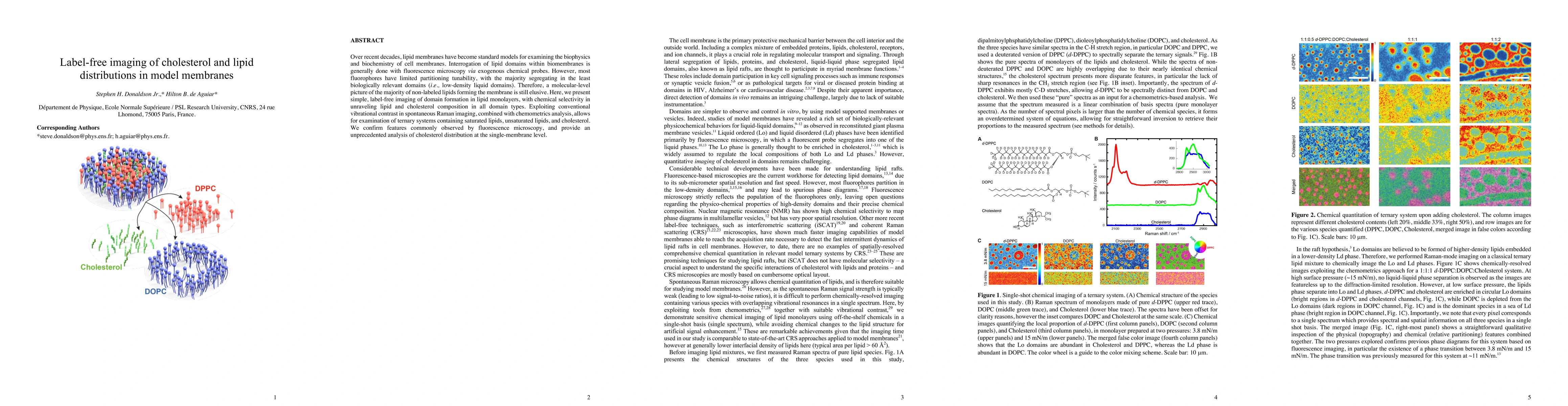
Over recent decades, lipid membranes have become standard models for examining the biophysics and biochemistry of cell membranes. Interrogation of lipid domains within biomembranes is generally done with fluorescence microscopy via exogenous chemical probes. However, most fluorophores have limited partitioning tunability, with the majority segregating in the least biologically relevant domains (i.e., low-density liquid domains). Therefore, a molecular-level picture of the majority of non-labeled lipids forming the membrane is still elusive. Here, we present simple, label-free imaging of domain formation in lipid monolayers, with chemical selectivity in unraveling lipid and cholesterol composition in all domain types. Exploiting conventional vibrational contrast in spontaneous Raman imaging, combined with chemometrics analysis, allows for examination of ternary systems containing saturated lipids, unsaturated lipids, and cholesterol. We confirm features commonly observed by fluorescence microscopy, and provide an unprecedented analysis of cholesterol distribution at the single-membrane level.
AI Key Findings
Get AI-generated insights about this paper's methodology, results, and significance.
Paper Details
PDF Preview
Key Terms
Citation Network
Current paper (gray), citations (green), references (blue)
Display is limited for performance on very large graphs.
Similar Papers
Found 4 papersDual effect of cholesterol on interfacial water dynamics in lipid membranes: Interplay between membrane packing and hydration
Kokoro Shikata, Kento Kasahara, Nozomi Morishita Watanabe et al.
| Title | Authors | Year | Actions |
|---|
Comments (0)