Label-free segmentation from cardiac ultrasound using self-supervised learning
Publication
Metrics
AI Quick Summary
This paper presents a self-supervised learning pipeline for segmenting cardiac chambers from ultrasound images, eliminating the need for manual annotations. The method achieved high accuracy in segmenting and measuring cardiac chambers, comparable to supervised learning and inter-clinician variation, and demonstrated strong correlation with MRI measurements.
Paper Preview
Abstract
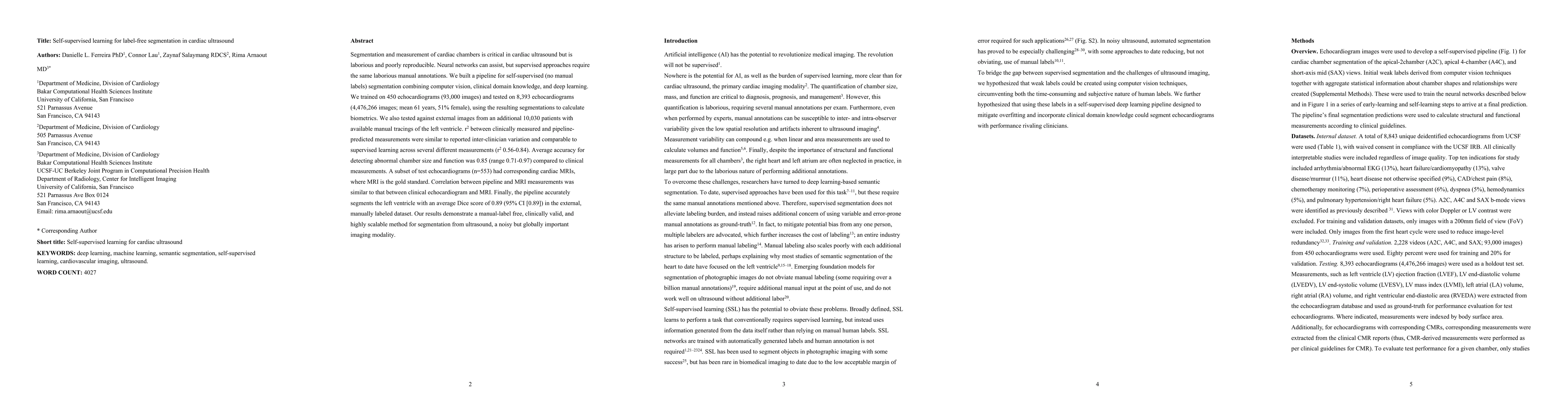
Segmentation and measurement of cardiac chambers is critical in cardiac ultrasound but is laborious and poorly reproducible. Neural networks can assist, but supervised approaches require the same laborious manual annotations. We built a pipeline for self-supervised (no manual labels) segmentation combining computer vision, clinical domain knowledge, and deep learning. We trained on 450 echocardiograms (93,000 images) and tested on 8,393 echocardiograms (4,476,266 images; mean 61 years, 51% female), using the resulting segmentations to calculate biometrics. We also tested against external images from an additional 10,030 patients with available manual tracings of the left ventricle. r2 between clinically measured and pipeline-predicted measurements were similar to reported inter-clinician variation and comparable to supervised learning across several different measurements (r2 0.56-0.84). Average accuracy for detecting abnormal chamber size and function was 0.85 (range 0.71-0.97) compared to clinical measurements. A subset of test echocardiograms (n=553) had corresponding cardiac MRIs, where MRI is the gold standard. Correlation between pipeline and MRI measurements was similar to that between clinical echocardiogram and MRI. Finally, the pipeline accurately segments the left ventricle with an average Dice score of 0.89 (95% CI [0.89]) in the external, manually labeled dataset. Our results demonstrate a manual-label free, clinically valid, and highly scalable method for segmentation from ultrasound, a noisy but globally important imaging modality.
AI Key Findings
Get AI-generated insights about this paper's methodology, results, significance, and more — seven facets brought into focus.
Impact
Paper Details
Authors
PDF Preview
Key Terms
Citation Network
Current paper (gray), citations (green), references (blue)
Display is limited for performance on very large graphs.
Discussion 0