Quick Answers
What methodology did the authors use?
The research employed a deep learning approach using convolutional neural networks (CNNs) to analyze radiographic images of the clavicle, elbow, and thumb. The models were trained on a large dataset with annotations for various pathologies, and their performance was evaluated using both inclusive and exclusive labeling strategies. More in Methodology →
What are the key results?
The inclusive model demonstrated higher sensitivity in detecting fractures and degenerative changes across all anatomical regions. — The exclusive model showed better specificity, particularly in distinguishing between benign and malignant lesions. More in Key Results →
Why is this work significant?
This research contributes to the field of medical imaging by providing a robust framework for automated diagnosis of bone pathologies, which can improve diagnostic accuracy and reduce workload for radiologists. More in Significance →
What are the main limitations?
The study relies on annotated datasets which may introduce bias if not representative of diverse patient populations. — Performance differences between internal and external datasets suggest potential generalizability issues. More in Limitations →
Abstract
Objectives: To evaluate GPT-4o's ability to extract diagnostic labels (with uncertainty) from free-text radiology reports and to test how these labels affect multi-label image classification of musculoskeletal radiographs. Methods: This retrospective study included radiography series of the clavicle (n=1,170), elbow (n=3,755), and thumb (n=1,978). After anonymization, GPT-4o filled out structured templates by indicating imaging findings as present ("true"), absent ("false"), or "uncertain." To assess the impact of label uncertainty, "uncertain" labels of the training and validation sets were automatically reassigned to "true" (inclusive) or "false" (exclusive). Label-image-pairs were used for multi-label classification using ResNet50. Label extraction accuracy was manually verified on internal (clavicle: n=233, elbow: n=745, thumb: n=393) and external test sets (n=300 for each). Performance was assessed using macro-averaged receiver operating characteristic (ROC) area under the curve (AUC), precision recall curves, sensitivity, specificity, and accuracy. AUCs were compared with the DeLong test. Results: Automatic extraction was correct in 98.6% (60,618 of 61,488) of labels in the test sets. Across anatomic regions, label-based model training yielded competitive performance measured by macro-averaged AUC values for inclusive (e.g., elbow: AUC=0.80 [range, 0.62-0.87]) and exclusive models (elbow: AUC=0.80 [range, 0.61-0.88]). Models generalized well on external datasets (elbow [inclusive]: AUC=0.79 [range, 0.61-0.87]; elbow [exclusive]: AUC=0.79 [range, 0.63-0.89]). No significant differences were observed across labeling strategies or datasets (p>=0.15). Conclusion: GPT-4o extracted labels from radiologic reports to train competitive multi-label classification models with high accuracy. Detected uncertainty in the radiologic reports did not influence the performance of these models.
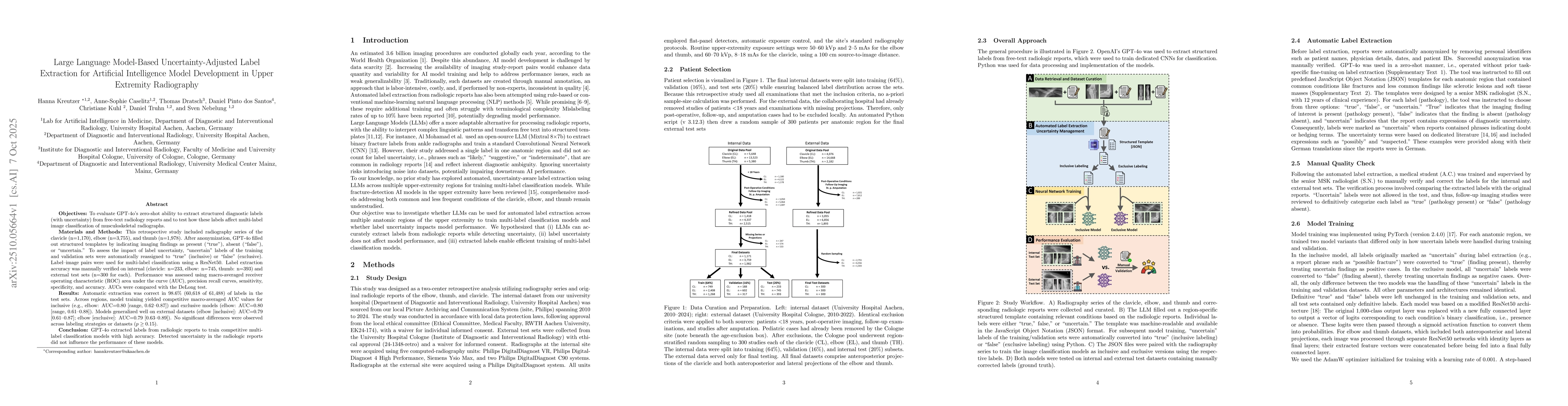
AI Key Findings
Generated Oct 09, 2025
Methodology — What approach did the authors take?
The research employed a deep learning approach using convolutional neural networks (CNNs) to analyze radiographic images of the clavicle, elbow, and thumb. The models were trained on a large dataset with annotations for various pathologies, and their performance was evaluated using both inclusive and exclusive labeling strategies.
Key Results — What are the main findings?
- The inclusive model demonstrated higher sensitivity in detecting fractures and degenerative changes across all anatomical regions.
- The exclusive model showed better specificity, particularly in distinguishing between benign and malignant lesions.
- Performance metrics varied significantly between internal and external validation datasets, highlighting the importance of dataset diversity.
Significance — Why does this research matter?
This research contributes to the field of medical imaging by providing a robust framework for automated diagnosis of bone pathologies, which can improve diagnostic accuracy and reduce workload for radiologists.
Technical Contribution — What is the technical contribution?
The technical contribution lies in the development of a dual-model framework that balances sensitivity and specificity through inclusive and exclusive labeling strategies, along with the comprehensive evaluation across multiple anatomical regions and datasets.
Novelty — What is new about this work?
This work introduces a novel approach to medical image analysis by combining both inclusive and exclusive labeling strategies in a single framework, offering a more nuanced understanding of model performance in diagnostic tasks.
Limitations — What are the limitations of this study?
- The study relies on annotated datasets which may introduce bias if not representative of diverse patient populations.
- Performance differences between internal and external datasets suggest potential generalizability issues.
Future Work — What did the authors propose for future work?
- Investigate the integration of multi-modal data (e.g., clinical history, lab results) with imaging data for more comprehensive diagnosis.
- Explore federated learning approaches to improve model generalization across different healthcare institutions.
- Develop explainable AI techniques to enhance clinical trust and adoption of the models.
Paper Details
How to Cite This Paper
@article{caselitz2025large,
title = {Large Language Model-Based Uncertainty-Adjusted Label Extraction for
Artificial Intelligence Model Development in Upper Extremity Radiography},
author = {Caselitz, Anne-Sophie and Kuhl, Christiane and Santos, Daniel Pinto dos and others},
year = {2025},
eprint = {2510.05664},
archivePrefix = {arXiv},
primaryClass = {cs.AI},
}
Caselitz, A., Kuhl, C., Santos, D., Truhn, D., Kreutzer, H., Nebelung, S., & Dratsch, T. (2025). Large Language Model-Based Uncertainty-Adjusted Label Extraction for
Artificial Intelligence Model Development in Upper Extremity Radiography. arXiv. https://arxiv.org/abs/2510.05664
Caselitz, Anne-Sophie, et al. "Large Language Model-Based Uncertainty-Adjusted Label Extraction for
Artificial Intelligence Model Development in Upper Extremity Radiography." arXiv, 2025, arxiv.org/abs/2510.05664.
PDF Preview
Similar Papers
Found 4 papersAccuracy of large language model-based artificial intelligence tools for equine topics.
Aldworth-Yang, S, Coleman, S J, O'Reilly, K et al.
An In-depth Survey of Large Language Model-based Artificial Intelligence Agents
Pengyu Zhao, Zijian Jin, Ning Cheng
GeneSUM: Large Language Model-based Gene Summary Extraction
Zhijian Chen, Chuan Hu, Min Wu et al.
Comments (0)