Multi-sequence Magnetic Resonance Imaging (MRI) offers remarkable
versatility, enabling the distinct visualization of different tissue types.
Nevertheless, the inherent heterogeneity among MRI sequences poses significant
challenges to the generalization capability of deep learning models. These
challenges undermine model performance when faced with varying acquisition
parameters, thereby severely restricting their clinical utility. In this study,
we present PRISM, a foundation model PRe-trained with large-scale
multI-Sequence MRI. We collected a total of 64 datasets from both public and
private sources, encompassing a wide range of whole-body anatomical structures,
with scans spanning diverse MRI sequences. Among them, 336,476 volumetric MRI
scans from 34 datasets (8 public and 26 private) were curated to construct the
largest multi-organ multi-sequence MRI pretraining corpus to date. We propose a
novel pretraining paradigm that disentangles anatomically invariant features
from sequence-specific variations in MRI, while preserving high-level semantic
representations. We established a benchmark comprising 44 downstream tasks,
including disease diagnosis, image segmentation, registration, progression
prediction, and report generation. These tasks were evaluated on 32 public
datasets and 5 private cohorts. PRISM consistently outperformed both
non-pretrained models and existing foundation models, achieving first-rank
results in 39 out of 44 downstream benchmarks with statistical significance
improvements. These results underscore its ability to learn robust and
generalizable representations across unseen data acquired under diverse MRI
protocols. PRISM provides a scalable framework for multi-sequence MRI analysis,
thereby enhancing the translational potential of AI in radiology. It delivers
consistent performance across diverse imaging protocols, reinforcing its
clinical applicability.
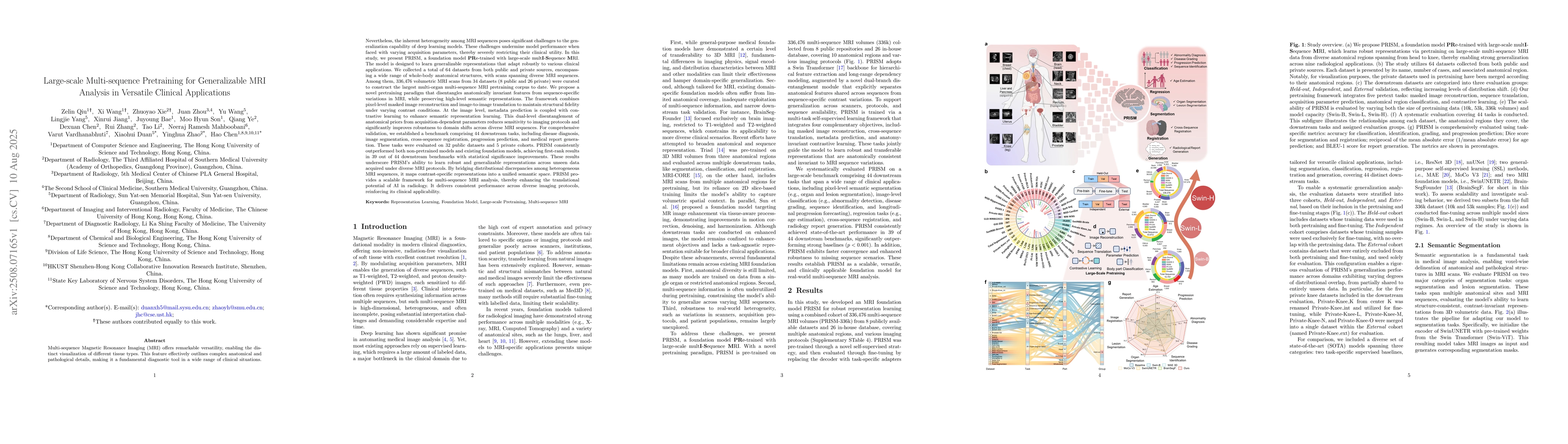
Discussion 0